
Recent Advances in Bioconjugated Transition Metal Complexes for Cancer Therapy

1
Dipartimento di Scienze Molecolari e Nanosistemi, Università Ca’ Foscari, Campus Scientifico, Via Torino 155, 30174 Venezia-Mestre, Italy
2
Dipartimento di Scienze Chimiche, Università degli Studi di Padova, Via Marzolo 1, 35131 Padova, Italy
*
Author to whom correspondence should be addressed.
Appl. Sci. 2023, 13(9), 5561; https://doi.org/10.3390/app13095561
Submission received: 7 April 2023
/
Revised: 26 April 2023
/
Accepted: 27 April 2023
/
Published: 29 April 2023
(This article belongs to the Special Issue Bioactive Compounds from Various Sources: Beneficial Effects and Technological Applications II)
Abstract
:The introduction of biologically relevant organic moieties in the coordination sphere of transition metal complexes has recently become a well-established strategy to increase the selectivity and biocompatibility of metallodrugs. In this review, the major advances achieved in this area of research in the last three years are described in detail. Particular attention is given to the metal complexes bearing the main biomolecules of life: carbohydrates, lipids, nucleotides, proteins and vitamins. Each paragraph summarizes the synthetic strategy employed to obtain the complexes of interest as well as the most interesting biological results obtained with these potential metallodrugs. Moreover, the structure–activity relationships observed by the different research groups are described and discussed, with the goal of suggesting to the reader the ligand/metal centre combinations that provide the most promising results in the fight against cancer. Some of the compounds examined in this review as well as other bioconjugated metal complexes published in recent decades exhibit interesting selectivity towards cancer cells over normal ones and a specific mode of action. These latter aspects are the basis of what is commonly known as anticancer target therapy.
1. Introduction
The human body is mainly composed of hydrogen, oxygen, carbon, sulphur, phosphorus and nitrogen (97.5% in total). These elements are the key ingredients of biomolecules, namely, carbohydrates, amino acids and proteins, nucleic acids and lipids. The remaining 2.5% of our body consists mostly of metals such as sodium, potassium, magnesium, calcium, manganese, iron, cobalt, copper, zinc and molybdenum. By virtue of their fundamental importance in the organism, they are considered “essential metals”. In fact, each of them plays a specific role in the metabolism such that a deficiency or an accumulation can cause serious diseases (e.g., anaemia in the case of iron deficiency) [1,2]. Modern analytical and computational techniques have allowed researchers to study in detail the exact role of essential metals in living systems and the interaction between metal ions/metal complexes and organic biomolecules [1,2]. Conversely, the role of the other metals present in the organism, which are considered “non-essentials”, is still debated.
As far as concerns the heavy metal ions, they are usually considered exogenous and toxic species [3]. However, following the famous assertion of Paracelso, “the right dose differentiates a poison from remedy”, heavy metal complexes are widely employed for the treatment of several diseases [4].
Historically, the use of metals in medicine started in the early 1900s with Salvarsan, an organoarsenic(III) compound with antimicrobial properties and particularly effective in the treatment of syphilis. Another milestone in the field of medicinal inorganic chemistry is the serendipitous discovery of the outstanding antitumor properties of cisplatin (cis-diaminodichloroplatinum(II)). The pioneering studies conducted by Rosenberg and the approval of this compound for clinical use in 1978 paved the way for the use of metal-based compounds for cancer therapy [5,6].
The high reactivity of metal complexes in the biological environment has made it difficult to predict their toxicity and therapeutic properties by computational methods (e.g., molecular docking), making the in vitro and in vivo screening steps indispensable [1,4,7,8]. Despite the huge number of metal-based compounds with interesting in vitro and in vivo anticancer properties, cisplatin and its second- and third-generation derivatives (e.g., carboplatin and oxaliplatin) remain the gold standards for the treatment of 50–70% of solid tumours. However, the non-negligible side-effects of platinated antineoplastic agents (e.g., nephrotoxicity, hepatotoxicity, neuro- and ototoxicity, cardiotoxicity, haematological and gastrointestinal toxicity) and their ineffectiveness towards some types of aggressive tumours (e.g., high-grade serous ovarian cancer, HGSOC) are the main limitations of this category of metallodrugs. All these issues as well as some suggestions for the design of new generations of metal-based anticancer agents are well described in recent reviews on this topic [6,9,10,11]. In particular, the investigation of complexes containing metals other than platinum and/or the development of efficient drug delivery systems (e.g., lipoplatin) are the two most prominent solutions proposed in these contributions [12].
The complexity of this area of research since the discovery of cisplatin has been well described in recent reviews from the Sodhi [13] and Sadler groups [14]. More specifically, they defined metallodrugs as unique metal–ligands combinations which, due to the wide range of processes that they can promote in the biological environment, can be considered challenging but extremely versatile and multitarget compounds. Their main mode of action depends on the activation mechanism, which is intimately related to the nature of metal centre and coordinated ligands, as well as the geometry and the overall stability of the complex in biological media. For this reason, the choice of biological assays and analytical techniques in the study of the mechanism of action of new metal-based drug candidates is extremely important to avoid false-positive or false-negative results [14].
The numerous studies conducted in the last 50 years on the activation mechanisms of metallodrugs in the biological environment have allowed researchers to classify metal complexes according to various criteria. A first subdivision is that between functional and structural compounds. In the case of structural compounds, the direct interaction of the metallodrug, in its original structure, with the biotarget is responsible for the therapeutic effect. Conversely, in the case of functional compounds, also known as prodrugs, the metal complex is not supposed to interact directly with the biotarget. More in detail, the metal complex undergoes modifications in the biological environment with respect to its original structure (e.g., release of one or more ligands), and only in this form it can interact with the main biotarget. In addition to their functional or structural behaviour, metal-based compounds can act as: (i) carriers for active ligands, (ii) catalysts or (iii) photoactivable species [15]. More recently, the classification of metallodrugs has been improved by adding more detailed classes such as: (i) bioconjugated metallodrugs, (ii) inhibitors of enzymes via substrate and metabolite mimics, (iii) redox-active derivatives [16], (iv) photoactivable compounds for photodynamic and photoactivated therapies [17,18,19,20], (v) metal complexes for delivery and release of pharmacologically active ligands, (vi) catalytic drugs [20,21], (vii) radiometals and radioactive agents for radio-imaging and radiotherapy [20,21] and (viii) magnetic resonance imaging (MRI) contrast agents [22].
In this vast scenario, some authors preferred to classify metal-based complexes of biological interest based on the type of metal centre, therapeutic effect and reactivity/stability in biological media [23,24,25,26,27,28,29,30]. In all these contributions, the design of heterobimetallic complexes and the conjugation of metal complexes to a bioinspired target moiety seem the most promising strategies to boost the efficacy of biologically active metal derivatives [31,32,33]. In particular, the new generations of metallodrugs are usually characterized by the presence of natural derivatives directly anchored to the metal centre and/or to the ancillary ligands [34]. The presence of a natural moiety is aimed at improving the biocompatibility of metallodrugs, thus reducing their side-effects and facilitating the drug delivery.
It should be remembered that with the term “natural”, we can consider all molecules that derive from living organisms present in nature. In this context, the most interesting bioconjugated metallodrugs contain plant derivatives such as coumarins and xanthines (e.g., caffeine) [35,36,37,38,39,40,41], food derivatives (e.g., polyphenols) [39,42] and classical biomolecules of life. Within this context, in 2019, Leung and Ma assembled a review dealing with metal complexes conjugated to natural products for cancer therapy [43]. In the same year, Hortelano, Estèvez-Braun and Heras fixed the state of the art of metal complexes with natural products from different biosynthetic pathways such as terpenes, curcumins, coumarins, steroids, naphto- and anthraquinones and tetracyclines [44].
Herein, we propose an overview of the recent advances in this vast area of research, with a special focus on metal-based anticancer agents bearing the essential biomolecules of life: carbohydrates, amino acids and proteins, lipids (steroid and hormones), nucleic acids and vitamins. For convenience, abbreviations for the cell lines and tumour types cited in the main text are reported in the final Appendix A.
2. Metal-Based Carbohydrate Drug Conjugates
Glycolysis is the first key step of the glucose metabolism. More specifically, one molecule of glucose is degraded, under anaerobic conditions, by a cascade of enzymatic reactions in two molecules of pyruvate with production of ATP (Adenosine Tri-Phosphate). In the absence of oxygen, pyruvate evolves into lactate or ethanol (fermentation). On the contrary, in the presence of oxygen, pyruvate is involved in the Krebs cycle for the respiration process. The latter, in conjunction with the oxidative phosphorylation, takes place in the mitochondria and produces a lot of energy compared to glycolysis.
Cancer cells, with their altered metabolism, do not exploit the traditional Krebs cycle/mitochondrial oxidative phosphorylation pathway as an energy source, even under aerobic conditions. The inefficient metabolism of cancer cells is based on a different mechanism, known as aerobic glycolysis, to produce the ATP necessary for their growth and proliferation. This phenomenon is called Warburg Effect, which is characterized by a higher uptake and consumption of glucose by cancer cells compared to normal ones [45]. As a consequence, the massive production of lactate from the aerobic glycolysis is responsible for the acidic pH of the tumour microenvironment.
Many studies have demonstrated the role of the Warburg Effect in the oncogenesis process as well as the ability of this abnormal glucose metabolism to induce the resistance of cancer cells towards classical chemotherapy. Additional details on how aerobic glycolysis, tumour microenvironment and signalling pathways affect the cancer therapy are well summarized by Fan in a recent review [46].
Taking advantage of the Warburg Effect, many research groups have employed glucose and its derivatives to develop analytical techniques aimed at identifying tumour masses (e.g., Positron Emission Tomography, PET) and/or to design selective anticancer drugs. In particular, the introduction of a glycosidic moiety in the chemical structure of a metal complex can, in theory, increase the uptake on cancer cells and the selectivity of the metallodrug. In addition, carbohydrate moieties can be used to improve the solubility of metal complexes in the biological media and can act as efficient drug delivery systems, especially in the case of cyclodextrins derivatives [47]. In this context, a comprehensive review dealing with carbohydrate–metal complexes with interesting anticancer properties was assembled by Minutolo and colleagues in 2020 [48], covering all the contributions published in this area until 2019.
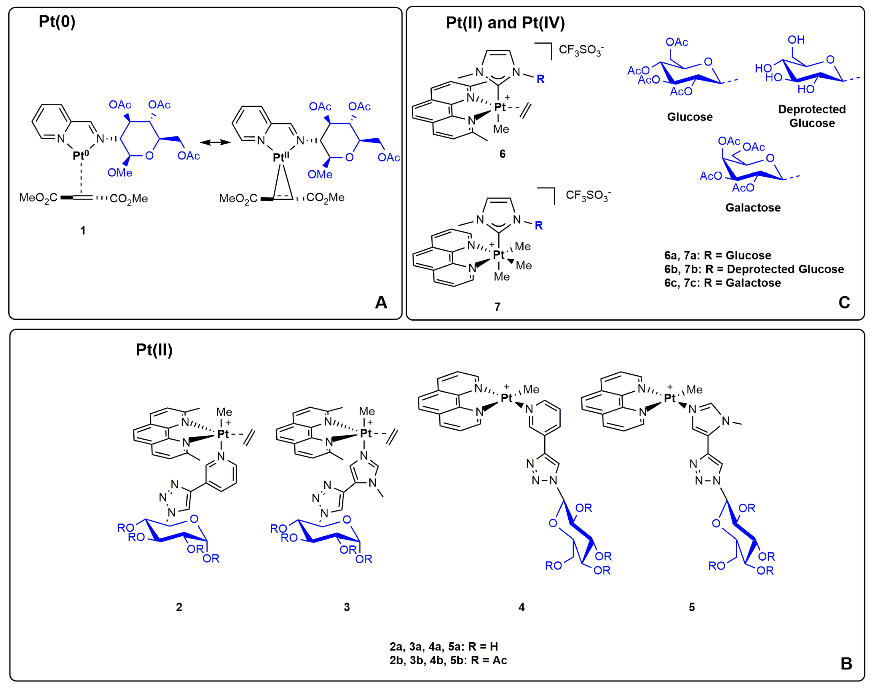
In the last three years, the Ruffo group investigated the synthesis and antitumor activity of a selection of platinum glycoconjugated complexes with different geometries and oxidation states of the metal centre [49,50,51]. In particular, the authors prepared in good yields and purity the platinum(0) complex (1) shown in Figure 1A, which bears a glycoconjugated 2-iminopyrydine and dimethylfumarate. This olefin ligand is currently used for the treatment of multiple sclerosis. The high degree of metal-to-olefin π-backdonation permits a parallelism with square planar Pt(II) complexes owing to the metallacyclopropane character of these formal Pt(0) species. The synthesized Pt(0) complex is water soluble, and its antiproliferative activity towards two tumoral (A431 and SVT2) and two non-tumoral (BALB/c3T3 and HaCaT) epithelial cell lines was evaluated by MTT assay after 48 h of incubation. The IC50 values indicate a superior cytotoxicity of this Pt(0) complex with respect to cisplatin in the SVT2 cancer cell line (IC50 = 61 µM). However, the similar IC50 values obtained on cancer and normal cells suggest a poor selective anticancer activity [49].
As we have mentioned in the introduction section, the stability and geometry of metal complexes are strictly related to their cytotoxic effect and mode of action. In this context, Ruffo and colleagues have reported the synthesis of five-coordinated trigonal bipyramidal Pt(II) complexes 2 and 3, which have shown high reactivity in physiological conditions (Figure 1B) [50]. In fact, the trans effect of the methyl group in axial position enables the easy substitution of pyridine or triazole ligands. On the contrary, square planar Pt(II) complexes 4 and 5 are very robust even in solutions with different ratios of physiological solution and DMSO. Notably, their lower reactivity can be explained by the presence of a weaker trans-labilizing ligand such as phenanthroline in trans to pyridine or triazole.
The biological results have shown a superior cytotoxicity on MCF-7 and A431 cancer cell lines of five-coordinated Pt(II) complexes 2 and 3 with respect to the four-coordinated Pt(II) complexes 4 and 5. The latter have shown DNA intercalation as confirmed by the ethidium bromide assay using calf-thymus DNA (ctDNA) as well as the activation of caspases 3 and 9 [50]. Moreover, complexes 2 and 3 exhibited a lower cytotoxicity towards H9c2 and HaCaT normal cell lines, thus suggesting a certain in vitro selectivity. Interestingly, a lower cytotoxicity was noticed in the case of glucose derivatives with respect to their acylglucose congeners.
Octahedral Pt(IV) complexes are considered excellent prodrugs and valid alternatives to classical square planar Pt(II) complexes. In Figure 1C, the general structures of Pt(II) and Pt(IV) complexes bearing carbohydrate-based NHC ligands are reported. The coordination sphere of platinum is completed with phenanthroline, methyl and ethylene (for Pt(II) species) ligands [51]. Pt(II)-NHC complexes 6a–c are sufficiently stable under physiological conditions. However, in the presence of stronger coordinating solvents (e.g., DMSO) the release of ethylene and phenanthroline was observed. On the contrary, Pt(IV)-NHC complexes 7a–c displayed a lower stability in aqueous solution, since the presence of their azolium salts was noticed by NMR studies. Additionally, in these categories of compounds, the key role of the type of glycosidic substituent used was confirmed. In particular, the presence of galactose renders these platinum complexes inactive towards MCF-7 and A431 cancer cells. At the same time, Pt(II)-NHC complexes exhibited a greater cytotoxicity compared to their Pt(IV)-NHC congeners (IC50 > 125 µM).
As expected, none of the compounds tested acts via DNA double-strain (dsDNA) intercalation. However, a direct interaction with both double-strain DNA and single-strain DNA was detected by ESI-MS. In addition, stability studies in the presence of a few selected proteins such as human serum albumin (HSA) and hen egg white lysozymes (HEWL) suggested a potential stable interaction [51].
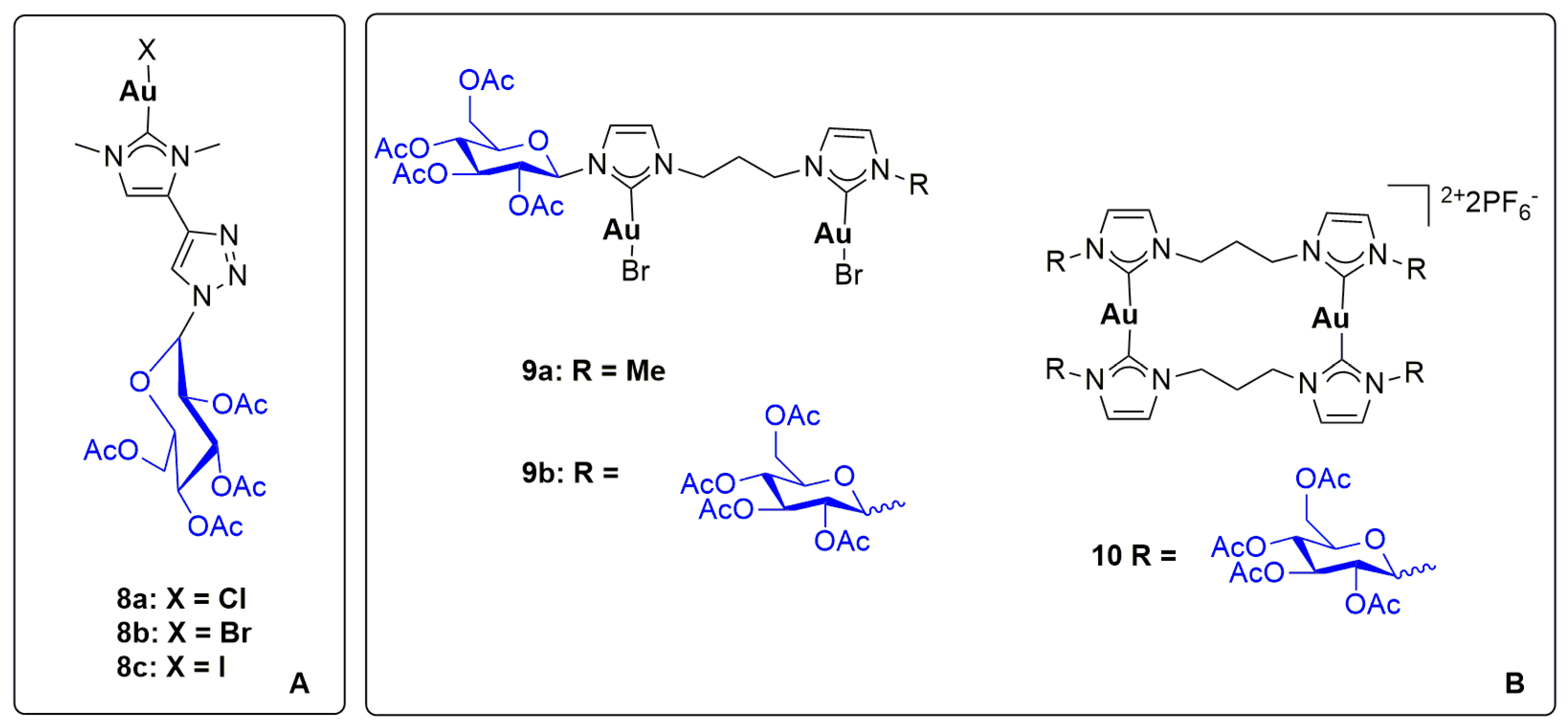
Moving to the gold chemistry, neutral Au(I) complexes bearing NHCs ligands are usually thioredoxin reductase inhibitors (TrxR), a selenoprotein belonging to the Trx system, which is located in the mitochondria. On the contrary, cationic Au(I) biscarbene complexes usually interact with DNA, in particular with G-quadruplex DNA structures [52,53,54,55,56]. Taking advantage of the easy click reaction, NHC ligands bearing a glycosidic triazole moiety can be synthesized and directly coordinated to gold [57]. More specifically, neutral gold-NHC complexes 8a–c, depicted in Figure 2A, present the same glycosidic-NHC but different halides. The compounds are quite stable in polar solvents (e.g., DMSO, DMSO-H2O and Acetone-H2O solutions). However, traces of cationic biscarbene species [Au(NHC)2]+ can be observed, especially in the NHC-Au-Cl complex.
The antiproliferative activity of complexes 8a–c was evaluated on A431 and SVT2 cancer cells and HaCaT and BALB/c3T3 normal cells. The obtained IC50 values are in the high micromolar range (35–100 µM), with the highest selectivity index in the case of complex 8c. The traditional circular dichroism and fluorescence techniques suggested that this class of complexes can alter the DNA conformation by intercalation, especially in the iodide derivative 8c. Moreover, crystallization with RNase A and egg white lysozyme proteins (HEWL) highlighted the ability of these gold-NHC complexes to bind specific protein residues.
As far as concerns the position and number of glycosidic moieties in a gold complex, Tubaro and colleagues have investigated a selection of dinuclear gold derivatives (Figure 2B) [58]. In particular, complexes 9a–b and 10 were synthesized and tested towards A431 and SVT2 cancer cells and HaCaT and BALB/c3T3 normal ones. Notably, the anticancer activity of the corresponding Au(III) complexes was not explored due to their poor stability in the physiological solution.
Unfortunately, both neutral complexes 9a–b exhibited high IC50 values (>100 µM). On the contrary, the biscationic complex 10 showed a moderate cytotoxicity (IC50 = 85 µM) and a certain selectivity towards cancer cells.
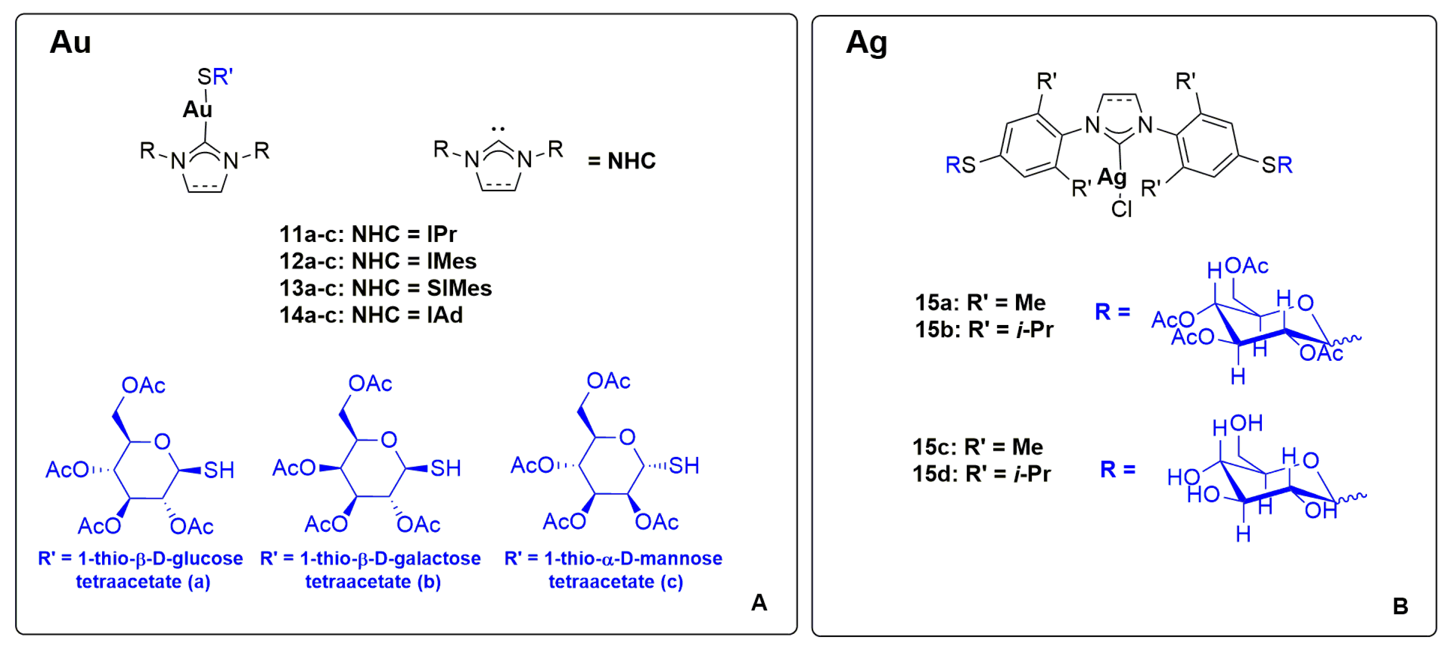
The well-known antiarthritic and antitumor properties of Auranofin have prompted many research groups to use thioglucosidic residues in the design of new promising metallodrugs. Taking advantage of the weak-base route [59], Nolan, Ott and Martin have recently reported the S-H activation of thiosugars under mild conditions, affording the desired gold complexes 11–14 with a wide scope of NHC ligands (Figure 3A) [60]. The synthesized complexes have been tested in relevant human cancer cell lines such as A549, HT-29 and MDAMB-231. All the tested compounds exhibited a remarkable anticancer activity, with IC50 values in the low micromolar or sub-micromolar range. Interestingly, the most promising results were obtained in the case of thioglucose tetraacetate as thiosugar.
As far as concerns the application of silver complexes in this area of research, Messaoudi, Lamaty and Gautier have developed a solvent-free procedure (via ball milling) for the preparation of Ag(I) complexes 15a–d (see Figure 3B) [61]. In particular, a selection of imidazolium salts bearing thiosugars in para position of the NHC-aryl substituents and Ag2O reacts smoothly in the ball mill apparatus, affording the desired complexes in high yields. The anticancer activity of these Ag-NHC complexes and their azolium precursors was evaluated on HCT 116 cancer cell line. The obtained results have demonstrated the superior antiproliferative activity of the silver complexes with respect to their azolium precursors. The highest activity was achieved with complex 15a [61].
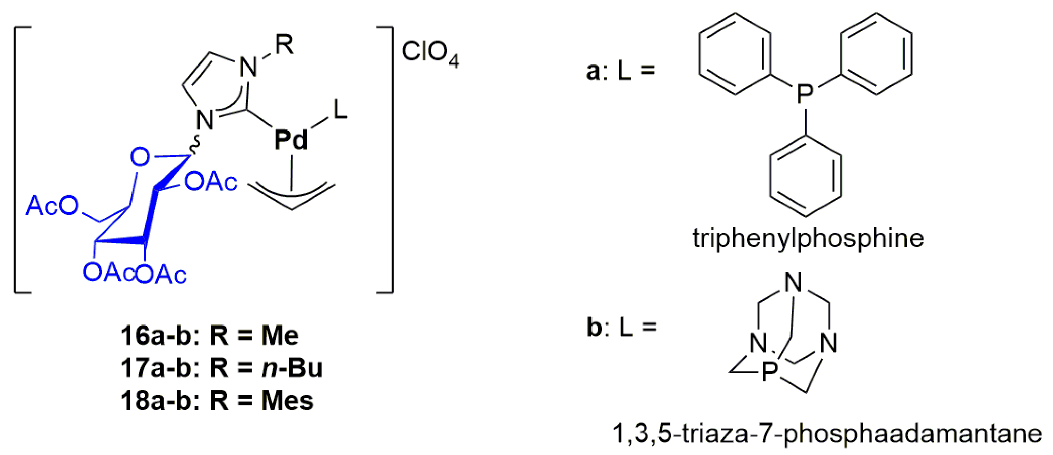
Organopalladium complexes have recently been considered as promising anticancer agents, owing to their remarkable in vitro, ex vivo and in vivo cytotoxicity even on cisplatin-resistant tumour models [62,63,64,65,66]. Recently contributions from our group have demonstrated the interesting anticancer properties of Pd(II)-allyl complexes bearing NHC and phosphine ligands [67]. More specifically, the use of PTA (1,3,5-triaza-7-phosphaadamantane) as phosphine ligand improves the solubility and stability of the Pd complexes in physiological conditions as well as their selectivity towards cancer cells. Moreover, this category of organopalladium derivatives seems to target mitochondria. With the aim of further improving their activity and selectivity, in 2020, we reported the use of glucosidic-NHCs as ancillary ligands in Pd(II)-allyl complexes. The desired complexes 16a–b, 17a–b and 18a–b were successfully obtained as a mixture of four diastereoisomers and then tested as potential anticancer agents (Figure 4).
It is noteworthy to mention that the very low IC50 values obtained on A2780, A2780cis, OVCAR-5, A549, A375, KURAMOCHI and OVCAR-3 cells have demonstrated the successful design of the compounds. More specifically, all IC50 values are in the low micromolar range and in all cases lower than cisplatin. The similar IC50 values obtained on A2780 and A2780cis (cisplatin-resistant clone) cell lines, suggest the different mechanism of action of these Pd-NHC complexes with respect to cisplatin. The highest cytotoxicity was observed in the case of complex 17a, whereas the lowest activity was observed in the case of complex 17b. Interestingly, the Pd complexes bearing PTA combine a good cytotoxicity towards cancer cells with a poor cytotoxicity towards normal ones (MRC-5 fibroblasts) [67].
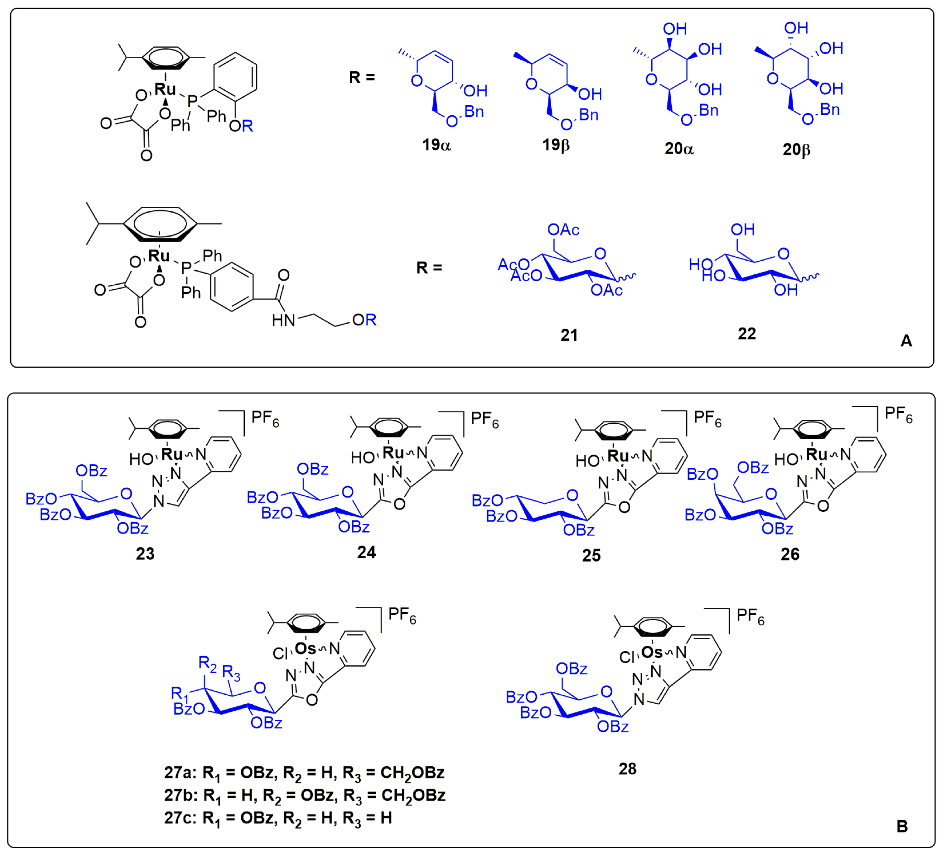
Moving to Group 8 of the Periodic Table, some interesting works dealing with the anticancer properties of iron, ruthenium and osmium complexes bearing glycosidic moieties have been published in the last three years [68,69,70]. In particular, ruthenium complexes are widely studied as chemotherapeutic agents as well as efficient photosensitizers for photodynamic therapy. Considering the glycosidic derivatives, Ru(II) p-cymene phosphane complexes depicted in Figure 5A have been recently synthesized, focusing on 2,3 unsaturated hexoses (compounds 19 and 20) and glucose derivatives (compounds 21 and 22). All the synthesized complexes have been tested towards ovarian and breast cancer cell lines (A2780, A2780cis and MCF7), showing a moderate-to-good antiproliferative activity only in the case of complexes 19α and 19β (IC50 = 2–20 µM). More specifically, the β isomer exhibited a superior activity compared to its α congener, but without a significant selectivity. In addition, these two Ru(II) complexes seem to act with different mechanisms of action. Complex 19β showed a remarkable accumulation of treated cells in S and G2/M phases. Conversely, complex 19α showed no alteration of the cell cycle. Although both complexes induced oxidative stress (ROS production), only complex 19β can activate Caspases 3/7 and damage the mitochondria membrane [68].
A panel of cationic half-sandwich Ru(II) p-cymene complexes have been prepared by Bokor and Bai (see Figure 5B), and the key role of the ancillary ligands in the cytotoxicity has been demonstrated [69]. In particular, complexes 23–26 showed IC50 values in the micromolar range (0.9–9 µM) on A2780 and ID8 ovarian cancer cell lines. Interestingly, the best candidates exhibited a very low cytotoxicity on normal cells (MRC-5 fibroblasts), thus demonstrating an interesting selectivity. Curiously, the use of deprotected sugars is sufficient to suppress the anticancer properties of the complexes. More in general, the increase in lipophilicity of the sugar moiety improves the cytostatic properties and induces a cooperative binding to unidentified targets with a remarkable ROS production.
With the aim of investigating the role of the metal centre, Bokor and Bai have reported the synthesis of osmium complexes 27 and 28 bearing the same ligands used in the previously mentioned ruthenium derivatives (Figure 5B). The biological tests have shown a lower cytotoxicity of osmium complexes compared to their ruthenium congeners [70].
Iron complexes, especially ferrocenyl derivatives, are well known as potent antimalarial, antifungal and anticancer agents. In this context, one of the most important examples is ferrocifen, which contains ferrocene and tamoxifen moieties in its structure.
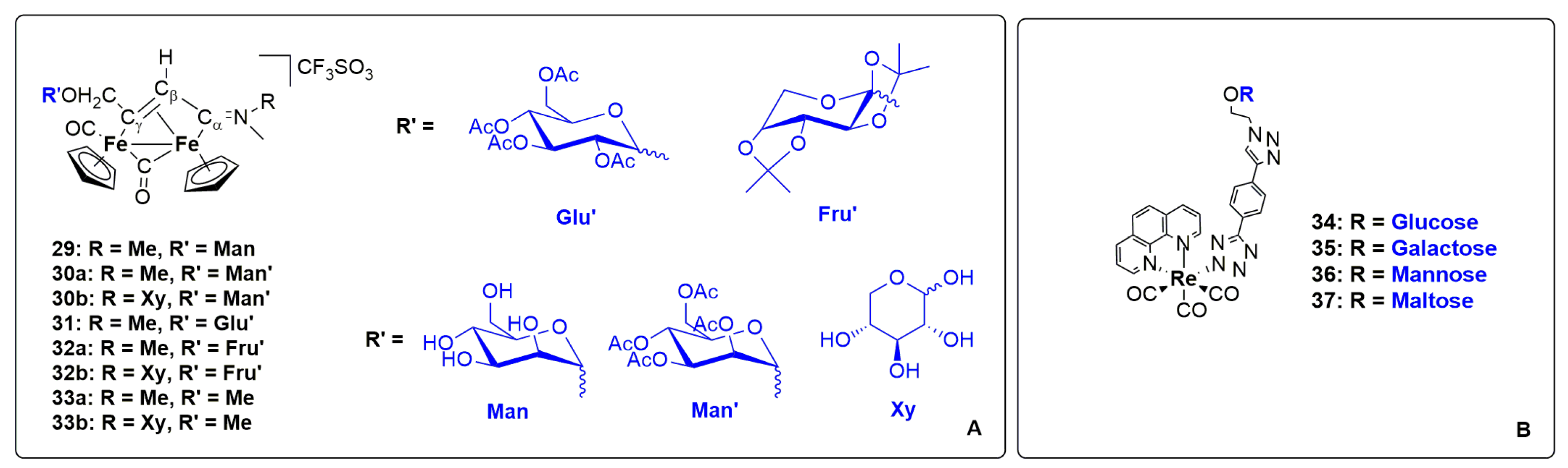
In this context, Marchetti, Di Bussolo and Gasser have reported in 2022 the preparation of interesting diiron vinilyminium complexes with glycosidic substituents (see Figure 6A) [71]. Most of the final complexes displayed a significant hydrophilicity, which is strongly influenced by the glycosidic portion employed. The anticancer activity of the selected compounds was evaluated on CT26, U87 and MCF-7 cancer cells, showing a good cytotoxicity only in the case of complexes 30b, 32b and 33b. However, these iron complexes exhibited a similar cytotoxicity also on RPE-1 normal cells.
Finally, Plush and Stagni have reported the synthesis and characterization of novel biocompatible Re(I) complexes 34–36 (Figure 6B) in which the glucosidic decoration has been easily obtained by click reaction [72]. The uptake of each complex in H9c2 cardiomyoblasts cells has been evaluated by ICP-MS. The results have confirmed the key role of lipophilicity in the cellular uptake by passive transport. In fact, complex 34 reported the highest uptake, in accordance with the highest lipophilic diffusion coefficient logD. Moreover, confocal laser scanning microscopy confirmed the ability of these complexes to be used as imaging agents with a light-induced toxicity biodistributed in endosomes/lysosomes and/or endoplasmic reticulum.
3. Metal-Based Protein, Peptide and Antibody Drug Conjugates
Specific sequences of amino acids form complex structures known as peptides and proteins, which are biomolecules involved in a wide range of organism vital functions. In recent times, amino acid derivatives have been investigated as anticancer agents, and in particular, poli-peptides [73] and antibodies [74,75] have been largely included in studies based on the immunotherapeutic approach. Furthermore, they can be conjugated with a metal-based scaffold in order to deliver the active moiety to the desired target taking advantage of their biocompatibility. Strategies to radiolabel bioconjugated metal complexes and to prepare photo-functional derivatives have been recently exhaustively reviewed [19,76,77,78,79,80,81,82]. In these contributions, the interaction between proteins and metallodrugs and the synthesis and characterization of the challenging bioconjugated drugs have been well described. Therefore, only the most relevant examples developed in the last three years will be illustrated in this section.
Among the conjugated proteins, HSA (human serum albumin) is certainly one of the most studied, being the most abundant blood protein, with important implications in transport processes and therefore for the pharmacokinetics of metallodrugs [83]. So far, only ruthenium and platinum-HSA conjugate were prepared with the aim of achieving the desired target and act after release of the active form [84].
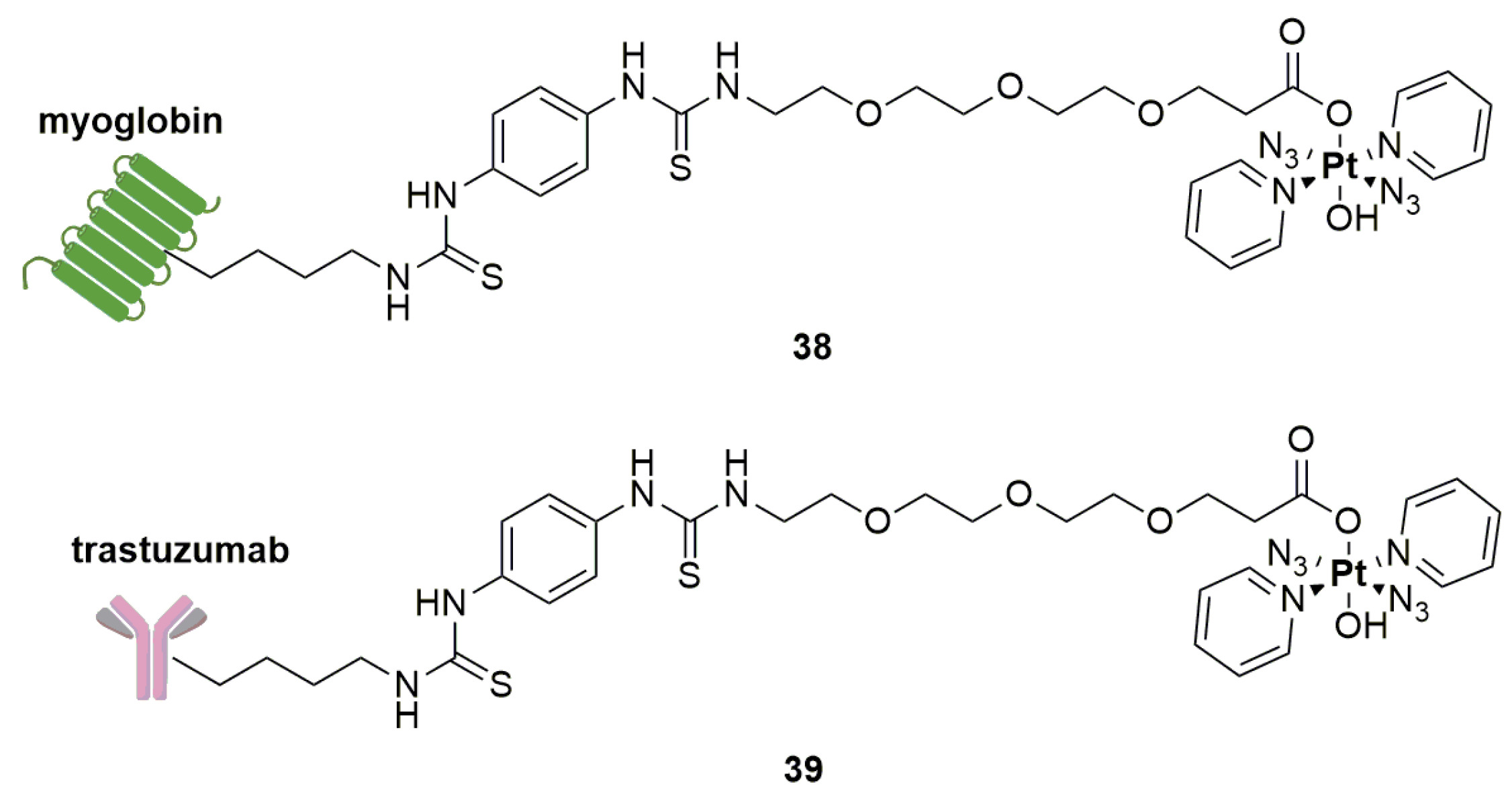
As already mentioned in the previous sections, octahedral Pt(IV) complexes are usually prodrugs [51]. The conjugation with a protein requires a water-compatible procedure that does not affect the protein conformation and allows the release of the cytotoxic payload once it reaches the tumour. Saldler and Imberti have described the photoactivable Pt(IV) azido compounds conjugated to myoglobin and trastuzumab antibody (complexes 38 and 39 in Figure 7) [85]. The conjugation with myoglobin is higher than with trastuzumab, and although both derivatives are stable in the dark, unfortunately, an extensive photodecomposition was observed during the preliminary tests.
The general structure of a bioconjugated metal peptide/antibody consists of three distinct building blocks: the metal, the linker and the peptide/antibody carrier [84]. Peptides conjugation is common, attractive and useful because of the cost-effective synthesis, biocompatibility, possibility of chemical optimization and the opportunity to take advantage of cell-penetrating peptides (CPP). CPP are sequences of 5–30 amino acids which form positively charged peptides able to cross the plasma membrane [82]. On the other hand, antibody–drug conjugates (ADC) represent a valid strategy for target therapy of blood and solid tumours. Usually, immunoglobulin or monoclonal antibody (mAb) produced by clones of a unique parent immune cell are employed. These systems, while ensuring a high selectivity, often present several problems for their synthesis, stability in plasma, efficient drug release and internalization. Furthermore, the cost of their synthesis is much higher than that of peptides [86,87]. However, in general, conjugation with metallodrugs is easier than with organic molecules, facilitating a reduction in production costs [78,84].
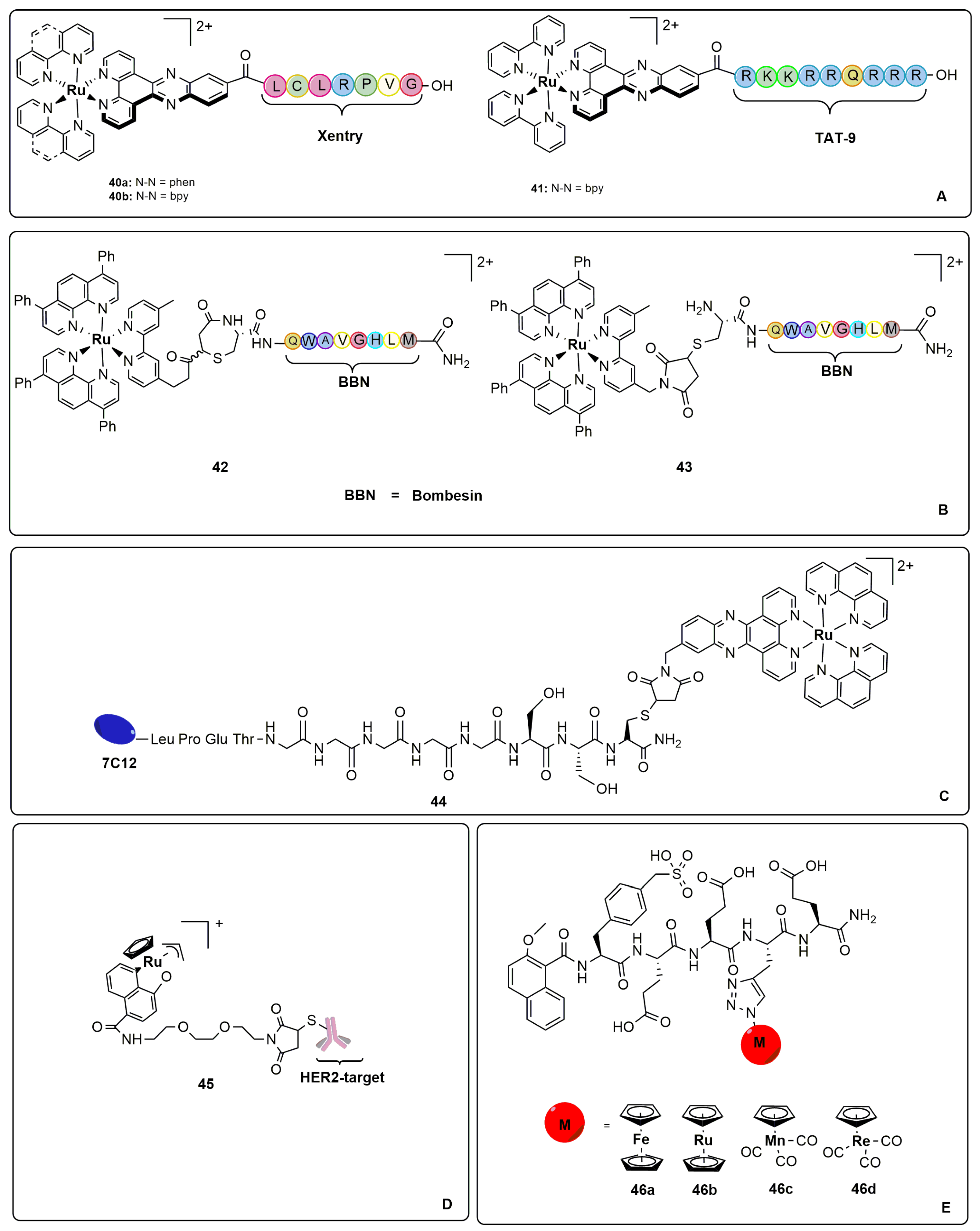
Ruthenium complexes, whose structures are reported in Figure 8, are interesting examples of bioconjugated metal peptides. In 2021, Metzler-Nolte reported the synthesis and DNA interaction studies of three cationic ruthenium polypyridyl dppz-CPP complexes (40 and 41, see Figure 8A) [88]. As known, they are usually investigated as luminescent probes in which the presence of the dppz ligand is crucial for DNA intercalation. Arginine TAT-9 and non-arginine Xentry neutral CPP have been chosen. As expected, all complexes interact with DNA, with a slight preference for TAT-9 derivatives, but unfortunately with minor differences with the non-bioconjugated metal derivatives. Furthermore, even the cellular uptake analysis has revealed the scarce impact of the use of CPPs, and no cytotoxicity towards HeLa and Hs-578T has been observed for either CPPs or the corresponding metal conjugated compounds (IC50 > 100 µM) [88].
In order to improve the panel of photosensitizers (PS) employed in photodynamic therapy (PDT), Gasser and Gois have proposed the conjugation of [Ru(bphen)2(dmbipy)]+ complexes with Bombesin peptide bearing a N-terminal Cys. The latter is known have high affinity with human gastrin-releasing peptide receptor (GRPR), which is overexpressed in many solid tumours [89]. Complexes 42 and 43, depicted in Figure 8B, were tested in PC-3, HT-29 and CT26 cancer cell lines in dark and light conditions, comparing the IC50 values to the non bioconjugated metal complexes. Bioconjugation has permitted us to maintain the cytotoxicity only in dark conditions (IC50 = 0.5–18 µM) in both complexes. Moreover, the general decreasing in cellular uptake was observed in the CT26 line, showing the highest accumulation of drug in lysosomes [89].
In complex 44 (see Figure 8C), ruthenium has been conjugated to the N7C12 nanobody, a specific binder to epidermal growth factor receptor (EGFR)-expressing cells [90]. A nanobody is a single chain domain, smaller than mAb, albeit with high stability, solubility, specificity, affinity and fast pharmacokinetics [82]. To encourage a specific site conjugation, a poly-glycine unit peptide was used as linker. The internalization of complex 44 into A431 cells is correlated to the capability of N7C12 to target EGFR in overexpressed cell lines after site-specific modification and is strongly dependent on the level of EGFR overexpression. The nanobody conjugation has not modified the photophysical properties of the metal complex, but no ROS production was detected, and consequently, no cytotoxicity in dark and in light conditions (IC50 > 25 µM due to complex precipitation) has been observed [90].
Compound 45 (Figure 8D) was reported in 2022 by Mao and Xia. It is an organometallic complex linked to an affibody, a small robust protein engineered to bind a large number of target proteins or peptides with high affinity [91]. Compound 45 bears a Human Epidermal Growth Factor Receptor 2 (HER2) target which is able to activate gemcitabine prodrug in situ via bio-orthogonal reaction and release the drug in proximity of the cell membrane. Moreover, owing to its bifunctional properties, bioconjugated complex 45 induced DNA damage in positive breast cancer cells, thus blocking their proliferation. The result has been confirmed even in spheroids and zebrafish in vitro and in vivo models, respectively [91].
Among compounds 46, depicted in Figure 8E, complex 46b, prepared by Gasser, Patra and Ferrari, is the most active of the first family of metallocene peptides with inhibitor properties towards cell division cycle 25 (CDC25) [92]. Despite the promising ex vivo preliminary results, chemical modifications or caging are necessary to improve the anticancer activity and cell membrane permeability [92].
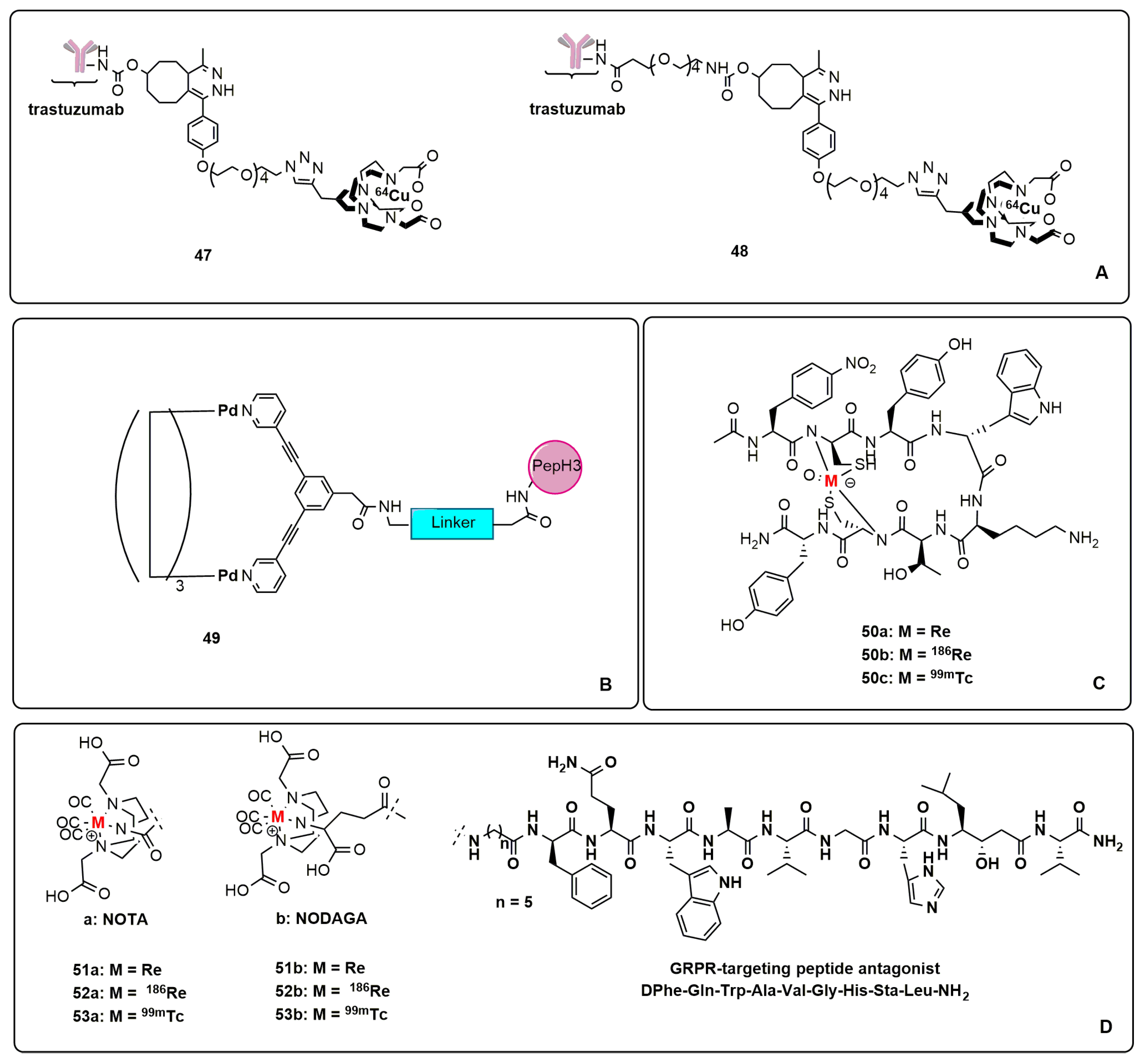
The immuno-positron emission technique (immune-PET) is a powerful tool to monitor the behaviour of radiolabelled antibodies in vivo in real time and it is therefore very useful to receive a rapid response in tumour diagnosis, immunotherapy and radioimmunotherapy. However, the structure of the bifunctional chelator (BFC) is crucial for the pharmacokinetics of the whole compound [93]. Usually, in bioconjugated complexes, BFC is coordinated to the radiometal and simultaneously bonded to the amino acid residue of the antibody. The most common BFCs for 64Cu metal centre are 1,4,7-triazacyclononane-1,4,7-triacetic acid (NOTA), 1,4,7,10-tetraazacyclododecane-1,4,7,10-tetraacetic acid (DOTA) and 1,4,8,11-tetraazacyclotetradecane-1,4,8,11- tetraacetic acid (TETA), but recently, they have been declared unstable in vivo. In 2021, Yoo and collaborators optimized a new category of propylene cross-bridged BFCs with a PEG-based linker to conjugate trastuzumab antibody in 64Cu complexes (47 and 48, Figure 9A) [94]. Both new platforms have shown in vivo stability and a higher uptake in HER2-positive cancer cell lines NIH3T6.7 and SKOV3 with respect to HER2-negative cells HEK293 and CT26 [94].
The strategy of self-assembling synthesis has been developed to prepare cationic [Pd2L4]4+ metallacages tethered to a blood–brain barrier (BBB)-penetrating peptide, PepH3, in order to overcome the well-known difficulty of ensuring drugs reach their brain targets. Complex 49, whose structure is depicted in Figure 9B, has been investigated by Casini, Correia and Mendes using bEnd.3 cells as an in vitro model to evaluate the ability of the metallacages to translocate the BBB [95]. In addition, the radiolabelling of the cage with [99mTcO4]−, the purification of the resulting radioactive cage and the assessment of its brain penetration properties were investigated in vivo. It is noteworthy to mention that the water-soluble system can cross the BBB only once conjugated to PepH3 moiety, and therefore, the radiolabelled [99mTcO4]− derivative presents a brain accumulation decidedly higher than that of the non-bioconjugated system [95].
Neuroendocrine tumours (NETs) are diagnosticated by imaging techniques such as PET and SPECT, which take advantage of the use of radiolabelled somatostatin analogues bearing radionuclides. Somatostatin receptors (SSTRs) are overexpressed in NETs, and they may be bound to the tracer that is able to internalize the agonist–receptor system into tumour cells. Radiolabelled tracers are usually designed using a bifunctional chelator introduced via the peptide N-terminus. More rarely, radiolabelled agents can be prepared via incorporation of the radiometal as an integral part of the peptide. The most employed radiometals are 99mTc and 186Re. In 2021, Hennkens and collaborators proposed the first 99mTc/186Re-cyclized SSTR antagonist peptide using the potent acetylated SSTR2 antagonist, Ac-4-NO2-Phe-c(DCys-Tyr-DTrp-Lys-Thr-Cys)-DTyr-NH2 [96]. The non-radioactive standard complex 50a, depicted in Figure 9C, has demonstrated good in vitro binding affinity on SSTR2-expressing AR42J cells (IC50 = 43 nM).
Among the 186Re/99mTc radiotracers 50b–c, only complex 50c exhibited good in vitro stability, and for this reason, it is a promising agent for diagnosis [97]. The same authors have also proposed GRPR-specific [99mTc/186Re]Tc/Re-tricarbonyl complexes using hydrophilic NOTA and NODAGA chelators conjugated via a 6-aminohexanoic acid linker (6-Ahx) to the powerful GRPR-targeting peptide antagonist D-Phe-Gln-Trp-Ala-Val-Gly-His-Sta-Leu-NH2 (Figure 9D). A high hydrophilicity and excellent in vitro and in vivo stability were observed for all complexes 51–53a–c. Moreover, a favourable pharmacokinetics in PC-3 murine tumour and a good receptor-mediated tumour uptake was noticed. Promising results have been reported by high-quality micro-SPECT/CT images for compound 53b. The reasons for its effectiveness could be attributed to the charge of the complex at pH = 7.4, although some further investigations are needed because some previous publications are at odds with this hypothesis. On the other hand, tumour retention should be improved in the case of rhenium complexes with the aim of reaching the therapeutic use [96].
4. Metal-Based Lipid and Nucleic Acid Drug Conjugates
Lipids are a class of hydrophobic molecules that include fatty acids, phospholipids, sterols, sphingolipids and prenol lipids. In general terms, they play an important role in cellular metabolism as well as in the mechanisms of drug absorption and internalization. Moreover, in the prodrug approach, lipids can be used as carriers [98]. The most studied are the steroids, especially in bioconjugated structures, because of their particularly stable and rigid framework. The favourable lipid solubility makes them suitable to penetrate the cell membrane and bind the specific receptors; for these reasons, they are largely employed in many therapeutic protocols, and many examples are reported in the literature relating to neoplastic diseases [99,100,101]. Since two very important reviews concerning steroid’s bioconjugation are published in 2021 [102] and 2022 [103], we will describe in this chapter only the most recent examples of bioconjugated lipid–metal complexes.
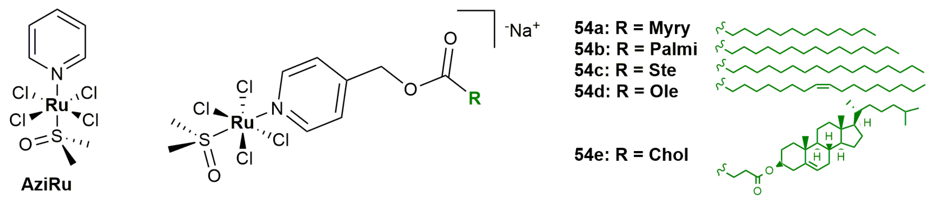
The encapsulation of drugs in lipid nanoparticles is a common and effective strategy to improve the drug delivery. In this context, Ru(III) derivatives synthesized by Montesarchio and co-workers are particularly worthy of mention. These octahedral complexes are characterized by the presence of a pyridyl ligand functionalized with a lipid chain, which facilitates the self-assembly in pseudo physiological conditions, with the resulting system that remains stable for hours [104]. More in detail, the behaviour of complexes 54a–e has been investigated and compared to the non-lipid functionalized pyridine precursor AziRu (Figure 10).
In this way, it was possible to observe that the lipidic chain protects the complex from the chloride ligand exchange, and this effect is particularly pronounced in the case of complexes 54c and 54e bearing the longest lipidic chain and the cholesterol scaffold, respectively. The lipophilicity of complexes 54b–d (logP ≈ 0.7) seems to increase their cytotoxicity towards MCF-7, MDAMB-231, HCT-116 and A375 cancer cells, whereas the same compounds are inactive in non-malignant cells (HaCaT and enterocytes). The high level of internalization in MDAMB-231 cells recorded for complex 54b (20 times higher with respect to AziRu precursor) may be one of the reasons of this increase in activity [104].
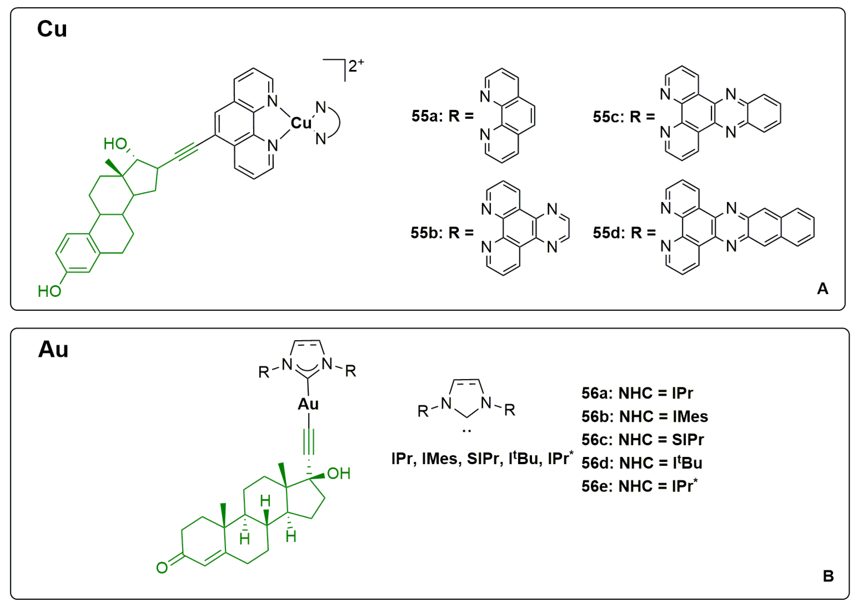
Steroids are important delivery vectors that can be used to target overexpressed oestrogen receptors (ER) cancer cells, such as those of breast, ovarian, colon and prostate tumours. The bioconjugation of oestrogen derivatives with the biocompatible copper metal centre has been studied by Erxleben and colleagues with the aim of understanding the interaction of phenanthroline ligands with DNA [105]. The oestradiol–copper complexes 55a–d (Figure 11A) are more effective than cisplatin against A431, MCF-7, 2008, A2780 ER+ and HCT-15 ER-cancer cells. The low and sub-micromolar activity has been confirmed even in 3D spheroids models, increasing with the lipophilicity of the phenanthroline ligands. The authors have shown that complex 55d is the most active, stimulates the production of ROS, and its uptake improves in a time-dependent manner. Overall, the activity of this class of compounds can be mainly ascribed to the electrostatic interaction (intercalation) with DNA, being scarcely able to establish covalent bonds [105].
Taking advantage of the well-known weak base route, [59] Nolan, Ott and collaborators synthesized linear gold(I) complexes bearing N-heterocyclic carbene (NHC) and ethisterone ligands (compounds 56a–e, depicted in Figure 11B) [106]. With the exception of compound 55e, all complexes inhibited the proliferation of A549, HT-29, MDA-MB-231 and MCF-7 cancer cells. Remarkably, minor differences in the structure of coordinated NHC involve significant differences in activity. The strategy to choose a specific agonist of progesterone receptor was a winning one, giving high uptake values in MCF-7 ER+ cells. Moreover, specific cytotoxicity assays, carried out simultaneously, administering ethisterone and complex 56a suggested that the coadministration might be competitive in the uptake process mediated by progesterone receptors [106].
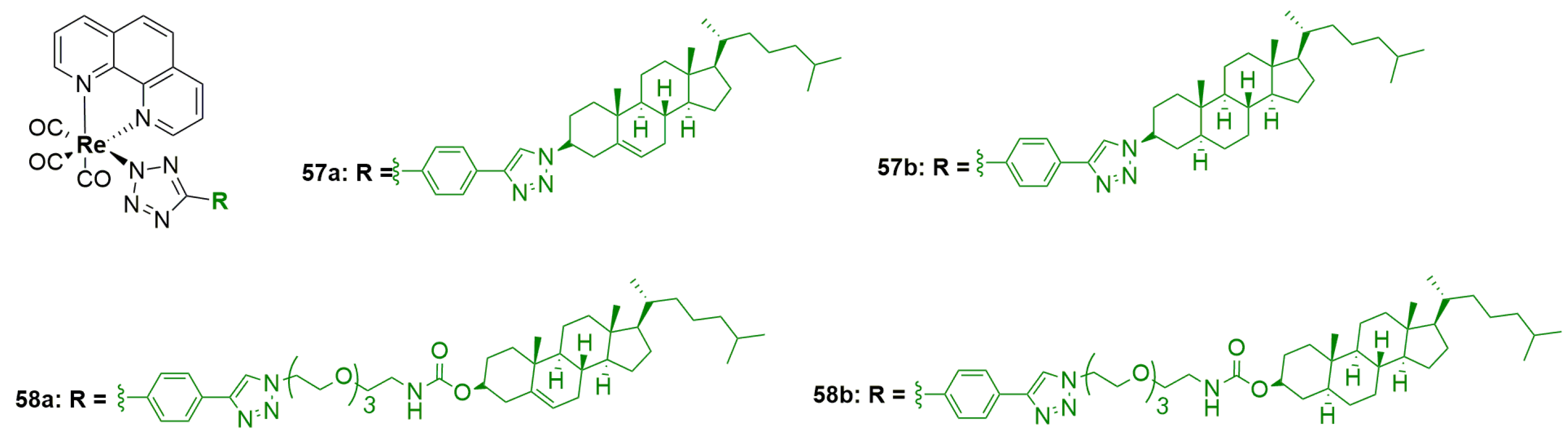
As written in previous sections, rhenium complexes are usually employed in fluorescence imaging techniques. Therefore, cholesterol and cholestanol rhenium complexes have been recently prepared by Brooks and colleagues [107].
Cholesterol is an essential multifunctional lipid involved in the control of eukaryotic cell membrane stability and permeability as well as a precursor to steroid hormones, bile acid and vitamin D3. Basically, its uptake takes place via endocytosis with low-density lipoproteins (LDL) to reach the endoplasmic reticulum. Cholesterol is present in high level in cancer cells, and for this reason, it is combined with rhenium for imaging applications, originating luminescent organometallic complexes which exhibit reduced photobleaching, large Stokes shift and long emission lifetimes compared to traditional organic fluorescent moieties. Adopting the classical click chemistry, it was possible to synthesize Re-cholesterol and cholestanol complexes 57a–b and their tetra ethylene glycol (TEG)-linked analogues 58a–b (see Figure 12). Unfortunately, for solubility issues, only complex 58b was investigated towards PC-3, LNCaP and 22Rv1 prostate cancer cells and PNT1a non-malignant cell line. The treatment with 5µM solution showed a higher uptake on cancer cells than non-tumoral ones. In particular, the complexes exhibited high cytotoxicity towards androgen-independent PC-3 cells, with the major accumulation in endosomes/lysosomes compartments [107].
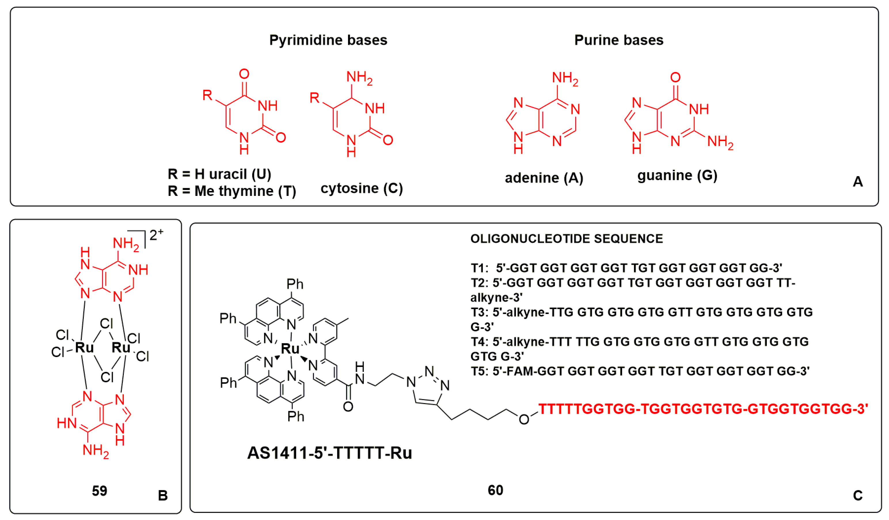
Nucleic acids are macromolecules dedicated to transport genetic information. DNA and RNA are constituted by nucleotides which are linked through a 3′,5′ phosphodiester bond. The main structural difference among them regards the nucleobases present in their sequences. More specifically, adenine, guanine, cytosine and thymine are involved in DNA sequences, while thymine is replaced by uracil in RNA (see Figure 13A). However, nucleic acids are usually not suitable for biological applications due to their sensitivity to nucleases, which causes a short half-life and therefore limits efficiency. For this reason, a wide range of synthetic nucleic acids, oligonucleotides, siRNA, miRNA, aptamer, plasmid nucleic acids, and purine and pyrimidine derivatives are usually employed [108,109,110].
Since a few recent reviews dealing with cisplatin [111] and other platinum complexes [112] bearing nucleic acid derivatives have been published, we have focused our attention on other metal centres. The main contributions in this research area involve the use of ruthenium complexes. In particular, in 2022, Martìnez-Lillo, Ribas and collaborators reported the first example of a dinuclear Ru(III) adenine protonated complex [113]. Compound 59, depicted in Figure 13B, was characterized from an electrochemical point of view and tested towards HCT-116 and AGS cell lines by MTT assay and cell cytometry after 48 and 72 h of treatment. This Ru(III) complex exhibits a specific cytotoxicity against AGS cancer cells in a time- and dose-dependent manner (IC50 = 23.25 µM after 72 h) displaying an apoptosis pathway in the highest concentration [113].
In the same year, Hollenstein, Gasser and colleagues have reported the synthesis and characterization of Ru(II) complexes bearing different oligonucleotides [114]. The real focus of their work was the investigation of the influence of the aptamer moiety. It should be remembered that aptamers are short oligonucleotides with high binding affinity to a specific target. Aptamers are usually used instead of an antibody because of their synthetic reproducibility, low cost and large-scale synthesis. AS1411 was chosen as the model aptamer thanks to the specific intercalating interaction with the DNA. Moreover, this aptamer can efficiently interact with nucleolin, a cell surface protein overexpressed in a wide range of cancer cells, including MCF-7 and DU145 models. Taking advantage of a click reaction, five Ru(II) complexes with T1–T5 oligonucleotides sequences were synthesized (Figure 13C). Interestingly, complex 60 displayed a promising Ru(II)-mediated photodynamic therapy towards MCF-7 cancer cells. It is noteworthy to mention that no toxicity was noticed on RPE-1 normal cells as well as no interference with G-quadruplex formation [114].
5. Metal-Based Vitamin Drug Conjugates
The last topic presented in this review concerns vitamin metal complexes.
Vitamins are essential organic molecules with a specific role in several phases of cell metabolism. Group B and C vitamins are water soluble, whereas those of A, D, E and K groups are lipid-soluble species. As usual, with platinum being the pioneer chemotherapeutic metal, its complexes are the most studied in this context too. In fact, bioconjugation of platinum prodrugs to vitamin E [115], its interactions with vitamin B [111] and catalytic properties of Pt-vitamin B12 complexes have already been extensively described in previous publications [116].
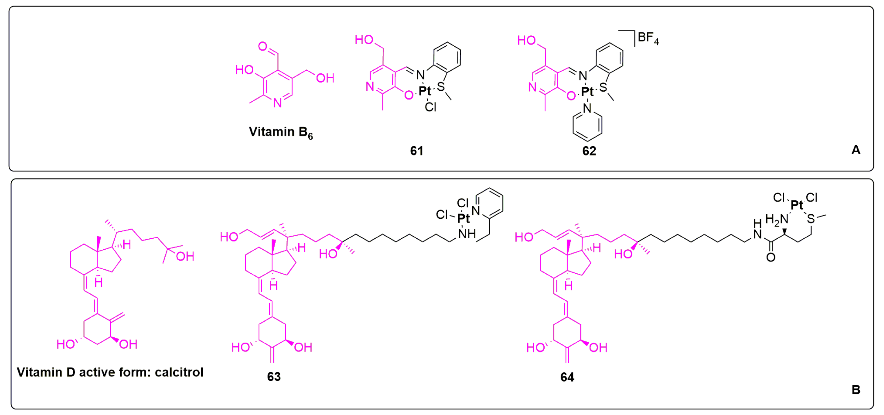
Pyridoxal, the natural form of vitamin B6, is a co-factor in enzymatic processes which is engaged in the metabolism of proteins, lipids and carbohydrates. Vitamin B6 mediates a wide range of biochemical reactions and influences cell growth. A few transition metal complexes based on pyridoxal exhibit strong antimicrobial and anticancer activities [111,117,118], and the first example of a Pt(II) derivative has been recently published by Rahman, Bhatti and co-workers [119]. More specifically, a vitamin B6 scaffold has been incorporated into a Schiff base ligand bearing ONS-donor atoms. Complexes 61 and 62, depicted in Figure 14A, induce follicular thyroid cancer (FTC) cell apoptosis showing a dose-dependent manner cytotoxicity. IC50 values have been determined by MTT assay and have been found comparable to cisplatin (IC50 = 9.6, 7.2 and 6.2 µM for 61, 62 and cisplatin, respectively). As free ligands are inactive, their coordination with the platinum centre is essential to promote their activity [119].
Calcitrol is the most bioactive form of Vitamin D3, a potent steroid hormone which regulates calcium and phosphate homeostasis. The gemini-type analogue of 19-norcalcitrol reported in Figure 14B has been coordinated to Pt(II) centres via two N donor atoms of 2-(2′-aminoethyl)pyridine moiety or via S and N donor atoms of the L-methionine fragment. Only complex 64 was a positive fit with the vitamin D receptor (VDR) binding assay. The cytotoxicity of complex 63 against CEM, MCF-7 and HeLa cancer cells is always in the low micromolar range (IC50 = 2.2–5.7 µM), outperforming the benchmark cisplatin. Remarkably, the anticancer activity of complex 64 is one order of magnitude lower than 63. Unfortunately, they are also both cytotoxic towards normal BJ fibroblast cells [120].
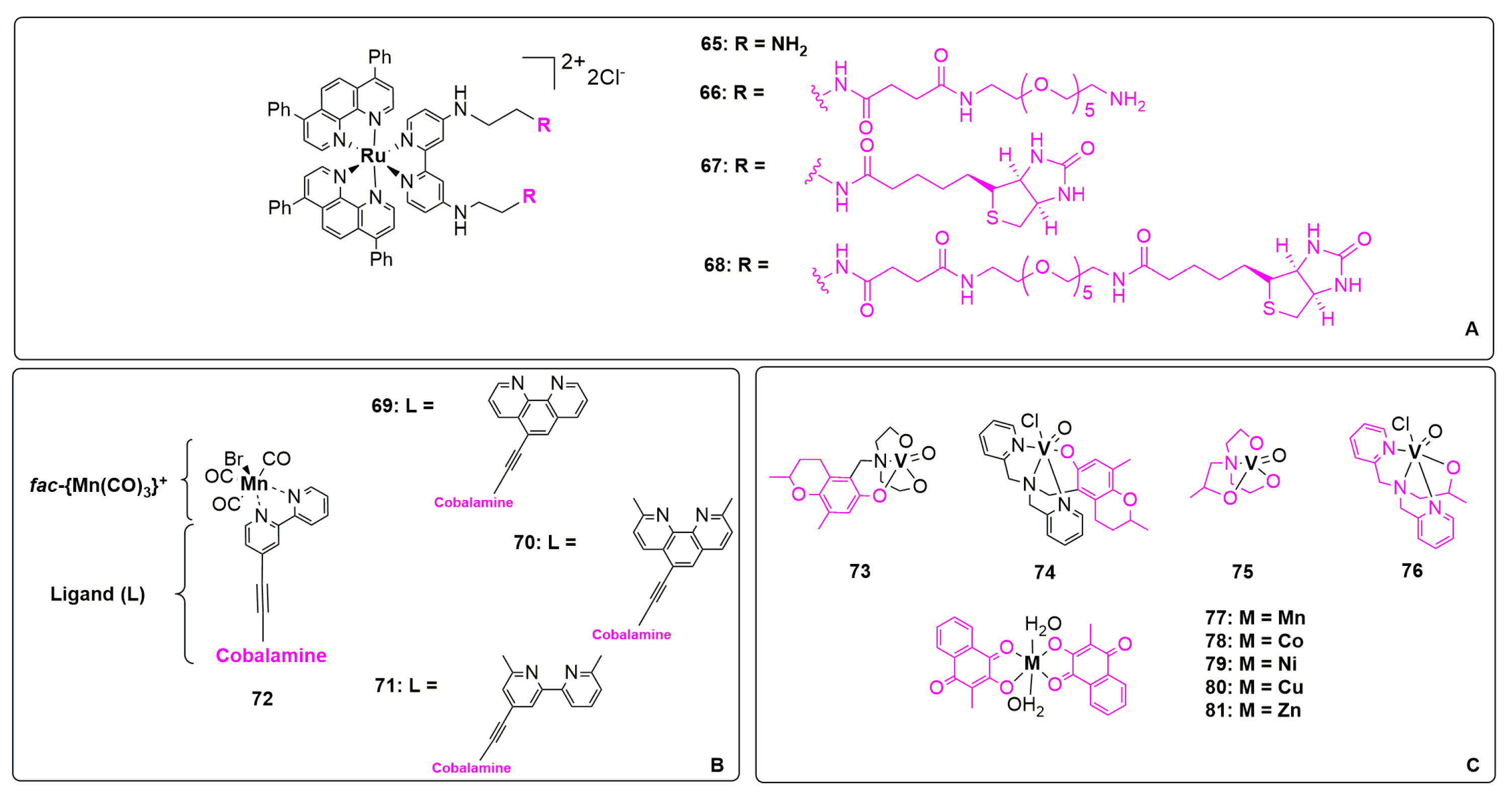
Ruthenium phenanthroline complexes including a vitamin moiety were reported by Gasser group in 2022 [121]. In particular, the influence of biotin (vitamin B8) was investigated in sodium-dependent multivitamin transport (SMVT)-overexpressed cell lines. SMVT are responsible for biotin uptake, which was therefore introduced in the coordination sphere of the complexes in order to enhance their uptake and cytotoxicity. Sometimes, biotin-based scaffolds are used even as nanocarriers in assembled systems. With the aim of improving the water solubility, pegylated derivatives were included in the study. The four complexes 65–68, depicted in Figure 15A, are generally hydrophilic but able to invert their affinity in physiological conditions, above all in the presence of plasma proteins, thanks to their propensity to form nanoparticles. It is noteworthy to mention that complex 67 showed the highest internalization in A549, CT26, HEK-293 and RPE-1 cells without showing significant cytotoxicity, due to its accumulation in the cell membrane. As a consequence, the common uptake by endocytosis observed for all four complexes has led to a final accumulation in lysosomes, excluding SMVT-dependent internalization. However, biotin-based complexes resulted in less toxicity than amino complexes, among which 65 and 68 exhibited phototoxicity after irradiation at 670 nm (with IC50 = 2.2–2.0 µM) [121].
Low concentrations of CO may favour cell proliferation while, on the contrary, high CO concentrations dramatically reduces the cell viability. CO molecule can be produced by heme-oxygenase enzymes or release in cell from CO-releasing molecules (CORMs). The coordination of CORMs on a metal centre may make them photoactivable. Taking advantage of the biocompatibility of manganese and transport function of vitamin B12 (cobalamin), Mn-CORMs have been prepared by Zobi, Rothen-Rutishauser and colleagues [122]. The chemical structure of these fac-{Mn(CO)3}+-B12 complexes 69–72 is reported in Figure 15B [122]. The authors have demonstrated that cobalamin is very important for the cellular uptake, acting as an active transporter through receptor-mediated endocytosis, whereas the internalization of non-cobalamin precursors happens by the passive diffusion. Since CO release is strictly cell-type dependent, A549, MCF-7, HT-29 cancer cells and 16HBE14o were used. Even if the role of the ligands in CO release is still unclear, it was noticed that cobalamin complexes are moderately cytotoxic before and after irradiation without evident differences. However, considering normal cells, coordinated CO plays a protective role in cell damage [122].
Vitamin E derivatives (tocopheryl and tocotryenyl) are known to selectively kill malignant cells mainly by ROS production. It is also well known that numerous vanadium complexes are able to promote a potent antitumor activity generally through the activation of apoptotic pathways and cell cycle arrest. Based on these assumptions, Keramidas and co-workers have designed some vanadium tocopherols derivatives as anticancer agents. Complexes 73–76, illustrated in Figure 15C, were tested towards cancer (LMS, U2OS and HeLa) and normal cell (MRC-5 and HEK-293) lines by MTT assay, showing all IC50 values in the micromolar and nanomolar range. The authors hypothesized that the synthesized complexes act in any case as ROS productors. It should be remembered that the capability of normal cells to tolerate a high ROS concentration is higher than that of cancer ones. Interestingly, the cell cycle is blocked in different manners independently of the type of complex administered. More in detail, in the LMS cell line, after 48 h, complex 73 causes cell cycle arrest in the S phase. Conversely, complex 74 causes cell cycle arrest in G2/M, and compound 75 shows a significant increase in the number of apoptotic cells (G0/G1 phase). Remarkably, complex 76 can act following different molecular pathways [123].
Finally, in a 2022 paper, Salunke-Gawali described a series of vitamin K3 metal derivatives [124]. Mn(II), Co(II), Ni(II), Cu (II) and Zn(II) phthiocol (2-hydroxy-3-methyl-1,4-naphthoquinone) complexes 77–81 (see Figure 15C) efficiently reduce the viability of MCF-7 and A549 cells and promote an apoptotic pathway. More in detail, the authors demonstrated that the cell cycle is arrested in the G1 phase in the case of breast cancer cells (MCF-7). In addition, mitochondria membrane potential is reduced, and the level of ROS is strongly increased by complex 79. In general, all compounds are able to induce DNA fragmentation, and notably, complex 80 inhibits topoisomerase II at 15 µM [124].
6. Conclusions
In conclusion, in this review, we have summarized the main advances obtained in the last three years in the field of bioconjugated metal complexes for cancer therapy. These recent results, in conjunction with those obtained in recent decades, have highlighted that the introduction of biologically relevant moieties in the coordination sphere of transition metal centres can improve the selectivity and biocompatibility of metallodrugs.
This approach was designed with the aim of promoting the transition from multimodal anticancer agents to metallodrugs with a specific biotarget and preferential accumulation on tumour cells.
In particular, most of the complexes described present ruthenium as the metal centre. Ruthenium complexes are in fact particularly suitable for bioconjugation by virtue of their high stability which significantly facilitates the purification of the final products. Such complexes have found extensive application in photodynamic therapy, with many examples currently in the clinical and preclinical stages.
For the other transition metals described in this review, the bioconjugation was carried out only after a careful choice of the ancillary ligands. The latter, especially phosphines, N-heterocyclic carbenes or chelating ligands, efficiently stabilize the metal centre, thus reducing or avoiding side-reactions that can occur during the introduction of bioactive moieties. Compounds with these transition metals (e.g., gold, palladium, copper, etc.) are studied as classical chemotherapeutic agents with the aim of replacing cisplatin and its derivatives in cancer therapy.
We strongly believe that the use of biomolecules such as carbohydrates, proteins, steroids, nucleotides and vitamins will continue to have a strong and growing interest both in the scientific community and in the pharmaceutical industry, with the common goal of consolidating what is known as anticancer target therapy.
In fact, the fight against cancer, which is the second most lethal class of pathologies worldwide, still requires enormous efforts for the development of new drugs as well as other efficient therapeutic and diagnostic approaches.
Author Contributions
Conceptualization, T.S., F.V. and E.B.; writing—original draft preparation, E.B.; writing—review and editing, T.S., F.V. and E.B.; supervision, T.S. and F.V. All authors have read and agreed to the published version of the manuscript.
Funding
This research received no external funding.
Institutional Review Board Statement
Not applicable.
Informed Consent Statement
Not applicable.
Data Availability Statement
The datasets generated and/or analysed during the present study are available from the corresponding author upon reasonable request.
Conflicts of Interest
The authors declare no conflict of interest.
Appendix A. Cell Lines
| Cancer cell lines | |
| MCF-7 | breast cancer |
| A431 | epidermoid carcinoma |
| SVT2 | murine fibroblasts BALB/c3T3 transformed with SV40 virus |
| A549 | lung adenocarcinoma |
| HT-29 | colon adenocarcinoma |
| MDAMB-231 | triple breast cancer |
| HCT-116 | colon carcinoma |
| A2780 | ovarian cancer |
| A2780cis | ovarian cancer cisplatin-resistant clone |
| A2780 | ovarian cancer |
| A2780cis | cisplatin-resistant ovarian cancer |
| OVCAR-3 | high-grade serous ovarian cancer |
| OVCAR-5 | high-grade serous ovarian cancer |
| KURAMOCHI | high-grade serous ovarian cancer |
| A375 | melanoma |
| ID8 | mouse ovarian surface epithelial cell line |
| CT26 | mouse colon cancer |
| U87 | glioblastoma cancer |
| HeLa | epithelial uterine cervix cancer |
| Hs-578T | breast cancer |
| PC-3 | prostate cancer |
| NIH3T6.7 | mouse fibroblast sarcoma sensitive |
| SKOV3 | high-grade serous ovarian cancer |
| AR42J | mouse pancreatic cancer |
| LNCaP | prostate cancer |
| 22Rv1 | prostate cancer |
| HCT-15 | colonrectal cancer |
| AGS | gastric adenocarcinoma |
| CEM | T lymphoblast |
| 16HBE14o | human bronchial epithelial cell line |
| LMS | Leiomyosarcoma |
| U2OS | osteosarcoma |
| Normal cell lines | |
| H9c2 | rat cardiomyoblast cells |
| HaCaT | human keratinocyte |
| BALB/c3T3 | immortalized murine fibroblasts |
| MRC-5 | lung fibroblast, normal cells |
| RPE-1 | retinoid mouse normal cells |
| PNT1a | human prostate normal cells |
| BJ | skin fibroblast |
| HEK293 | human kidney tissue |
References
- Stewart, T.J. Across the Spectrum: Integrating Multidimensional Metal Analytics for In Situ Metallomic Imaging. Metallomics 2019, 11, 29–49. [Google Scholar] [CrossRef] [PubMed] [Green Version]
- Bartnicka, J.J.; Blower, P.J. Insights into Trace Metal Metabolism in Health and Disease from PET: “PET Metallomics”. J. Nucl. Med. 2018, 59, 1355–1359. [Google Scholar] [CrossRef] [PubMed] [Green Version]
- Balali-Mood, M.; Naseri, K.; Tahergorabi, Z.; Khazdair, M.R.; Sadeghi, M. Toxic Mechanisms of Five Heavy Metals: Mercury, Lead, Chromium, Cadmium, and Arsenic. Front. Pharmacol. 2021, 12, 227–246. [Google Scholar] [CrossRef] [PubMed]
- Egorova, K.S.; Ananikov, V.P. Toxicity of Metal Compounds: Knowledge and Myths. Organometallics 2017, 36, 4071–4090. [Google Scholar] [CrossRef] [Green Version]
- Zhou, Y.; Ip, T.K.Y.; Zhang, Q.; Li, H.; Sun, H. Metal Complexes as Drugs and Therapeutic Agents. In Comprehensive Coordination Chemistry III; Elsevier: Amsterdam, The Netherlands, 2021; Volume 1–9, pp. 680–705. [Google Scholar]
- Ghosh, S. Cisplatin: The First Metal Based Anticancer Drug. Bioorg. Chem. 2019, 88, 102925. [Google Scholar] [CrossRef]
- Eric, A.G. Blomme Toxicology Strategies for Drug Discovery: Present and Future. Chem. Res. Toxicol. 2016, 29, 473–504. [Google Scholar]
- Deng, H.; Lei, Q.; Wu, Y.; He, Y.; Li, W. Activity-Based Protein Profiling: Recent Advances in Medicinal Chemistry. Eur. J. Med. Chem. 2020, 191, 112151. [Google Scholar] [CrossRef]
- Ferraro, M.G.; Piccolo, M.; Misso, G.; Santamaria, R.; Irace, C. Bioactivity and Development of Small Non-Platinum Metal-Based Chemotherapeutics. Pharmaceutics 2022, 14, 954. [Google Scholar] [CrossRef]
- Stankovic, J.S.K.; Selakovic, D.; Mihailovic, V.; Rosic, G. Antioxidant Supplementation in the Treatment of Neurotoxicity Induced by Platinum-Based Chemotherapeutics—A Review. Int. J. Mol. Sci. 2020, 21, 7753. [Google Scholar] [CrossRef]
- Valente, A.; Podolski-Renić, A.; Poetsch, I.; Filipović, N.; López, Ó.; Turel, I.; Heffeter, P. Metal- and Metalloid-Based Compounds to Target and Reverse Cancer Multidrug Resistance. Drug Resist. Updates 2021, 58, 100778. [Google Scholar] [CrossRef]
- Stathopoulos, G.P.; Boulikas, T. Lipoplatin Formulation Review Article. J. Drug Deliv. 2012, 2012, 581363. [Google Scholar] [CrossRef] [Green Version]
- Sodhi, R.K.; Paul, S. Metal Complexes in Medicine: An Overview and Update from Drug Design Perspective. Cancer Ther. Oncol. Int. J. 2019, 14, 25–32. [Google Scholar] [CrossRef]
- Anthony, E.J.; Bolitho, E.M.; Bridgewater, H.E.; Carter, O.W.L.; Donnelly, J.M.; Imberti, C.; Lant, E.C.; Lermyte, F.; Needham, R.J.; Palau, M.; et al. Metallodrugs Are Unique: Opportunities and Challenges of Discovery and Development. Chem. Sci. 2020, 11, 12888–12917. [Google Scholar] [CrossRef]
- Gianferrara, T.; Bratsos, I.; Alessio, E. A Categorization of Metal Anticancer Compounds Based on Their Mode of Action. Dalton Trans. 2009, 37, 7588–7598. [Google Scholar] [CrossRef]
- Amarsy, I.; Papot, S.; Gasser, G. Stimuli-Responsive Metal Complexes for Biomedical Applications. Angew. Chem. Int. Ed. 2022, 61, e202205900. [Google Scholar] [CrossRef]
- Gourdon, L.; Cariou, K.; Gasser, G. Phototherapeutic Anticancer Strategies with First-Row Transition Metal Complexes: A Critical Review. Chem. Soc. Rev. 2022, 51, 1167–1195. [Google Scholar] [CrossRef]
- Mfouo-Tynga, I.S.; Dias, L.D.; Inada, N.M.; Kurachi, C. Features of Third Generation Photosensitizers Used in Anticancer Photodynamic Therapy: Review. Photodiagnosis Photodyn. Ther. 2021, 34, 102091. [Google Scholar] [CrossRef]
- Lee, L.C.C.; Huang, L.; Leung, P.K.K.; Lo, K.K.W. Recent Development of Photofunctional Transition Metal−Peptide Conjugates for Bioimaging and Therapeutic Applications. Eur. J. Inorg. Chem. 2022, 35, e202200455. [Google Scholar] [CrossRef]
- Madec, H.; Figueiredo, F.; Cariou, K.; Roland, S.; Sollogoub, M.; Gasser, G. Metal Complexes for Catalytic and Photocatalytic Reactions in Living Cells and Organisms. Chem. Sci. 2023, 14, 409–442. [Google Scholar] [CrossRef]
- Konč, J.; Sabatino, V.; Jiménez-Moreno, E.; Latocheski, E.; Pérez, L.R.; Day, J.; Domingos, J.B.; Bernardes, G.J.L. Controlled In-Cell Generation of Active Palladium(0) Species for Bioorthogonal Decaging. Angew. Chem.—Int. Ed. 2022, 134, e202113519. [Google Scholar] [CrossRef]
- Boros, E.; Dyson, P.J.; Gasser, G. Classification of Metal-Based Drugs According to Their Mechanisms of Action. Chem 2020, 6, 41–60. [Google Scholar] [CrossRef] [PubMed]
- Lozhkin, B.; Ward, T.R. Bioorthogonal Strategies for the In Vivo Synthesis or Release of Drugs. Bioorg. Med. Chem. 2021, 45, 116310. [Google Scholar] [CrossRef] [PubMed]
- Martínez-Calvo, M.; Mascareñas, J.L. Organometallic Catalysis in Biological Media and Living Settings. Coord. Chem. Rev. 2018, 359, 57–79. [Google Scholar] [CrossRef]
- Rebelein, J.G.; Ward, T.R. In Vivo Catalyzed New-to-Nature Reactions. Curr. Opin. Biotechnol. 2018, 53, 106–114. [Google Scholar] [CrossRef] [Green Version]
- Chang, T.C.; Tanaka, K. In Vivo Organic Synthesis by Metal Catalysts. Bioorg. Med. Chem. 2021, 46, 116353. [Google Scholar] [CrossRef]
- Wang, J.; Wang, X.; Fan, X.; Chen, P.R. Unleashing the Power of Bond Cleavage Chemistry in Living Systems. ACS Cent. Sci. 2021, 7, 929–943. [Google Scholar] [CrossRef]
- Soldevila-Barreda, J.J.; Romero-Canelón, I.; Habtemariam, A.; Sadler, P.J. Transfer Hydrogenation Catalysis in Cells as a New Approach to Anticancer Drug Design. Nat. Commun. 2015, 6, 6582. [Google Scholar] [CrossRef] [Green Version]
- Nguyen, D.P.; Nguyen, H.T.H.; Do, L.H. Tools and Methods for Investigating Synthetic Metal-Catalyzed Reactions in Living Cells. ACS Catal 2021, 11, 5148–5165. [Google Scholar] [CrossRef]
- Destito, P.; Vidal, C.; López, F.; Mascareñas, J.L. Transition Metal-Promoted Reactions in Aqueous Media and Biological Settings. Chem. Eur. J. 2021, 27, 4789–4816. [Google Scholar] [CrossRef]
- Giorgi, E.; Binacchi, F.; Marotta, C.; Cirri, D.; Gabbiani, C.; Hangan, C.; Lucaciu, R.L.; Giorgi, E.; Binacchi, F.; Marotta, C.; et al. Highlights of New Strategies to Increase the Efficacy of Transition Metal Complexes for Cancer Treatments. Molecules 2022, 28, 273. [Google Scholar] [CrossRef]
- Shumi, G.; Desalegn, T.; Demissie, T.B.; Ramachandran, V.P.; Eswaramoorthy, R. Metal Complexes in Target-Specific Anticancer Therapy: Recent Trends and Challenges. J. Chem. 2022, 2022, 9261683. [Google Scholar] [CrossRef]
- Paprocka, R.; Wiese-Szadkowska, M.; Janciauskiene, S.; Kosmalski, T.; Kulik, M.; Helmin-Basa, A. Latest Developments in Metal Complexes as Anticancer Agents. Coord. Chem. Rev. 2022, 452, 214307. [Google Scholar] [CrossRef]
- Atanasov, A.G.; Zotchev, S.B.; Dirsch, V.M.; Orhan, I.E.; Banach, M.; Rollinger, J.M.; Barreca, D.; Weckwerth, W.; Bauer, R.; Bayer, E.A.; et al. Natural Products in Drug Discovery: Advances and Opportunities. Nat. Rev. Drug Discov. 2021, 20, 200–216. [Google Scholar] [CrossRef]
- Babahan-Bircan, I.; Emirdağ, S.; Özmen, A.; Abbak, M.; Ujam, O.T.; Demirkaya, I.; Günay, M.E. A New Hybrid Ligand and Its Metal Complexes from a Natural Plant (Styrax officinalis) Bearing Egonol, Thiosemicarbazone and Oxime Units, and Their Anti-Cancer Activities. Appl. Organomet. Chem. 2022, 36, e6784. [Google Scholar] [CrossRef]
- Scattolin, T.; Caligiuri, I.; Canovese, L.; Demitri, N.; Gambari, R.; Lampronti, I.; Rizzolio, F.; Santo, C.; Visentin, F. Synthesis of New Allyl Palladium Complexes Bearing Purine-Based NHC Ligands with Antiproliferative and Proapoptotic Activities on Human Ovarian Cancer Cell Lines. Dalton Trans. 2018, 47, 13616–13630. [Google Scholar] [CrossRef]
- Scattolin, T.; Giust, S.; Bergamini, P.; Caligiuri, I.; Canovese, L.; Demitri, N.; Gambari, R.; Lampronti, I.; Rizzolio, F.; Visentin, F. Palladacyclopentadienyl Complexes Bearing Purine-Based N-Heterocyclic Carbenes: A New Class of Promising Antiproliferative Agents against Human Ovarian Cancer. Appl. Organomet. Chem. 2019, 33, e4902. [Google Scholar] [CrossRef]
- Scattolin, T.; Pangerc, N.; Lampronti, I.; Tupini, C.; Gambari, R.; Marvelli, L.; Rizzolio, F.; Demitri, N.; Canovese, L.; Visentin, F. Palladium (0) Olefin Complexes Bearing Purine-Based N-Heterocyclic Carbenes and 1,3,5-Triaza-7-Phosphaadamantane (PTA): Synthesis, Characterization and Antiproliferative Activity toward Human Ovarian Cancer Cell Lines. J. Organomet. Chem. 2019, 899, 120857. [Google Scholar] [CrossRef]
- Balewski, Ł.; Szulta, S.; Jalińska, A.; Kornicka, A. A Mini-Review: Recent Advances in Coumarin-Metal Complexes with Biological Properties. Front. Chem. 2021, 9, 781779. [Google Scholar] [CrossRef]
- Halter, O.; Plenio, H. Fluorescent Dyes in Organometallic Chemistry: Coumarin-Tagged NHC–Metal Complexes. Eur. J. Inorg. Chem. 2018, 2018, 2935–2943. [Google Scholar] [CrossRef]
- Patil, S.A.; Kandathil, V.; Sobha, A.; Somappa, S.B.; Feldman, M.R.; Bugarin, A.; Patil, S.A. Comprehensive Review on Medicinal Applications of Coumarin-Derived Imine–Metal Complexes. Molecules 2022, 27, 5220. [Google Scholar] [CrossRef]
- Kim, J.; Lee, K.; Nam, Y.S. Metal-Polyphenol Complexes as Versatile Building Blocks for Functional Biomaterials. Biotechnol. Bioprocess Eng. 2021, 26, 689–707. [Google Scholar] [CrossRef]
- Ma, D.L.; Wu, C.; Cheng, S.S.; Lee, F.W.; Han, Q.B.; Leung, C.H. Development of Natural Product-Conjugated Metal Complexes as Cancer Therapies. Int. J. Mol. Sci. 2019, 20, 341. [Google Scholar] [CrossRef] [PubMed] [Green Version]
- Heras, B.L.; Amesty, Á.; Estévez-Braun, A.; Hortelano, S. Metal Complexes of Natural Product Like-Compounds with Antitumor Activity. Anticancer Agents Med. Chem. 2019, 19, 48–65. [Google Scholar] [CrossRef] [PubMed]
- Liberti, M.V.; Locasale, J.W. The Warburg Effect: How Does It Benefit Cancer Cells? Trends Biochem. Sci. 2016, 41, 211–218. [Google Scholar] [CrossRef] [PubMed] [Green Version]
- Liu, C.; Jin, Y.; Fan, Z. The Mechanism of Warburg Effect-Induced Chemoresistance in Cancer. Front. Oncol. 2021, 11, 3408. [Google Scholar] [CrossRef]
- Franconetti, A.; López, Ó.; Fernandez-Bolanos, J.G. Carbohydrates: Potential Sweet Tools Against Cancer. Curr. Med. Chem. 2020, 27, 1206–1242. [Google Scholar] [CrossRef]
- Bononi, G.; Iacopini, D.; Cicio, G.; Di Pietro, S.; Granchi, C.; Di Bussolo, V.; Minutolo, F. Glycoconjugated Metal Complexes as Cancer Diagnostic and Therapeutic Agents. ChemMedChem 2021, 16, 30–64. [Google Scholar] [CrossRef]
- Annunziata, A.; Cucciolito, M.E.; Imbimbo, P.; Silipo, A.; Ruffo, F. A Hydrophilic Olefin Pt(0) Complex Containing a Glucoconjugated 2-Iminopyridine Ligand: Synthesis, Characterization, Stereochemistry and Biological Activity. Inorg. Chim. Acta 2021, 516, 120092. [Google Scholar] [CrossRef]
- Annunziata, A.; Cucciolito, M.E.; Esposito, R.; Ferraro, G.; Monti, D.M.; Merlino, A.; Ruffo, F. Five-Coordinate Platinum(II) Compounds as Potential Anticancer Agents. Eur. J. Inorg. Chem. 2020, 11–12, 918–929. [Google Scholar] [CrossRef]
- Annunziata, A.; Amoresano, A.; Cucciolito, M.E.; Esposito, R.; Ferraro, G.; Iacobucci, I.; Imbimbo, P.; Lucignano, R.; Melchiorre, M.; Monti, M.; et al. Pt(II) versus Pt(IV) in Carbene Glycoconjugate Antitumor Agents: Minimal Structural Variations and Great Performance Changes. Inorg. Chem. 2020, 59, 4002–4014. [Google Scholar] [CrossRef]
- Casini, A.; Thomas, S.R. The Beauty of Gold: Knowledge of Mechanisms Leads to Different Applications of Organogold Compounds in Medicine and Catalysis. Chem. Lett. 2021, 50, 1516–1522. [Google Scholar] [CrossRef]
- Bindoli, A.; Rigobello, M.P.; Scutari, G.; Gabbiani, C.; Casini, A.; Messori, L. Thioredoxin Reductase: A Target for Gold Compounds Acting as Potential Anticancer Drugs. Coord. Chem. Rev. 2009, 253, 1692–1707. [Google Scholar] [CrossRef]
- Gratteri, P.; Massai, L.; Michelucci, E.; Rigo, R.; Messori, L.; Cinellu, M.A.; Musetti, C.; Sissi, C.; Bazzicalupi, C. Interactions of Selected Gold(III) Complexes with DNA G Quadruplexes. Dalton Trans. 2015, 44, 3633. [Google Scholar] [CrossRef]
- Wragg, D.; Ndreia De Almeida, A.; Iccardo Bonsignore, R.; Kühn, E.; Leoni, S.; Casini, A. G-Quadruplexes On the Mechanism of Gold/NHC Compounds Binding to DNAG-Quadruplexes:Combined Metadynamics and Biophysical Methods. Angew. Chem. 2018, 130, 14732–14736. [Google Scholar] [CrossRef]
- Bayrakdar, T.A.C.A.; Scattolin, T.; Ma, X.; Nolan, S.P. Dinuclear Gold(I) Complexes: From Bonding to Applications. Chem. Soc. Rev. 2020, 49, 7044–7100. [Google Scholar] [CrossRef]
- Annunziata, A.; Ferraro, G.; Cucciolito, M.E.; Imbimbo, P.; Tuzi, A.; Monti, D.M.; Merlino, A.; Ruffo, F. Halo Complexes of Gold(I) Containing Glycoconjugate Carbene Ligands: Synthesis, Characterization, Cytotoxicity and Interaction with Proteins and DNA Model Systems. Dalton Trans. 2022, 51, 10475–10485. [Google Scholar] [CrossRef]
- Tresin, F.; Stoppa, V.; Baron, M.; Biffis, A.; Annunziata, A.; D’Elia, L.; Monti, D.M.; Ruffo, F.; Roverso, M.; Sgarbossa, P.; et al. Synthesis and Biological Studies on Dinuclear Gold(I) Complexes with Di-(N-Heterocyclic Carbene) Ligands Functionalized with Carbohydrates. Molecules 2020, 25, 3850. [Google Scholar] [CrossRef]
- Scattolin, T.; Nolan, S.P. Synthetic Routes to Late Transition Metal–NHC Complexes. Trends Chem. 2020, 2, 721–736. [Google Scholar] [CrossRef]
- Safir Filho, M.; Scattolin, T.; Dao, P.; Tzouras, N.V.; Benhida, R.; Saab, M.; Van Hecke, K.; Lippmann, P.; Martin, A.R.; Ott, I.; et al. Straightforward Synthetic Route to Gold(I)-Thiolato Glycoconjugate Complexes Bearing NHC Ligands (NHC = N-Heterocyclic Carbene) and Their Promising Anticancer Activity. New J. Chem. 2021, 45, 9995–10001. [Google Scholar] [CrossRef]
- Ryzhakov, D.; Beillard, A.; Le Bideau, F.; Al-Shuaeeb, R.A.A.; Alami, M.; Bantreil, X.; Bonnemoy, A.; Gautier, A.; Lamaty, F.; Messaoudi, S. Azoliums and Ag(I)-N-Heterocyclic Carbene Thioglycosides: Synthesis, Reactivity and Bioactivity. Eur. J. Org. Chem. 2022, 9, e202101499. [Google Scholar] [CrossRef]
- Kapdi, A.R.; Fairlamb, I.J.S. Anti-Cancer Palladium Complexes: A Focus on PdX2L2, Palladacycles and Related Complexes. Chem. Soc. Rev. 2014, 43, 4751–4777. [Google Scholar] [CrossRef] [PubMed]
- Scattolin, T.; Voloshkin, V.A.; Visentin, F.; Nolan, S.P. A Critical Review of Palladium Organometallic Anticancer Agents. Cell Rep. Phys. Sci. 2021, 2, 100446. [Google Scholar] [CrossRef]
- Scattolin, T.; Pessotto, I.; Cavarzerani, E.; Canzonieri, V.; Orian, L.; Demitri, N.; Schmidt, C.; Casini, A.; Bortolamiol, E.; Visentin, F.; et al. Indenyl and Allyl Palladate Complexes Bearing N-Heterocyclic Carbene Ligands: An Easily Accessible Class of New Anticancer Drug Candidates. Eur. J. Inorg. Chem. 2022, 2, e2022000103. [Google Scholar] [CrossRef]
- Scattolin, T.; Bortolamiol, E.; Palazzolo, S.; Caligiuri, I.; Perin, T.; Canzonieri, V.; Demitri, N.; Rizzolio, F.; Cavallo, L.; Dereli, B.; et al. The Anticancer Activity of an Air-Stable Pd(I)-NHC (NHC = N-Heterocyclic Carbene) Dimer. Chem. Commun. 1223, 56, 12238. [Google Scholar] [CrossRef] [PubMed]
- Scattolin, T.; Bortolamiol, E.; Visentin, F.; Palazzolo, S.; Caligiuri, I.; Perin, T.; Canzonieri, V.; Demitri, N.; Rizzolio, F.; Togni, A. Palladium(II)-H3-Allyl Complexes Bearing N-Trifluoromethyl N-Heterocyclic Carbenes: A New Generation of Anticancer Agents That Restrain the Growth of High-Grade Serous Ovarian Cancer Tumoroids. Chem. Eur. J. 2020, 26, 11868–11876. [Google Scholar] [CrossRef]
- Scattolin, T.; Bortolamiol, E.; Rizzolio, F.; Demitri, N.; Visentin, F. Allyl Palladium Complexes Bearing Carbohydrate-Based N-Heterocyclic Carbenes: Anticancer Agents for Selective and Potent In Vitro Cytotoxicity. Appl. Organomet. Chem. 2020, 34, e5876. [Google Scholar] [CrossRef]
- Iacopini, D.; Vančo, J.; Di Pietro, S.; Bordoni, V.; Zacchini, S.; Marchetti, F.; Dvořák, Z.; Malina, T.; Biancalana, L.; Trávníček, Z.; et al. New Glycoconjugation Strategies for Ruthenium(II) Arene Complexes via Phosphane Ligands and Assessment of Their Antiproliferative Activity. Bioorg. Chem. 2022, 126, 105901. [Google Scholar] [CrossRef]
- Kacsir, I.; Sipos, A.; Ujlaki, G.; Buglyó, P.; Somsák, L.; Bai, P.; Bokor, É. Ruthenium Half-Sandwich Type Complexes with Bidentate Monosaccharide Ligands Show Antineoplastic Activity in Ovarian Cancer Cell Models through Reactive Oxygen Species Production. Int. J. Mol. Sci. 2021, 22, 10454. [Google Scholar] [CrossRef]
- Kacsir, I.; Sipos, A.; Bényei, A.; Janka, E.; Buglyó, P.; Somsák, L.; Bai, P.; Bokor, É. Reactive Oxygen Species Production Is Responsible for Antineoplastic Activity of Osmium, Ruthenium, Iridium and Rhodium Half-Sandwich Type Complexes with Bidentate Glycosyl Heterocyclic Ligands in Various Cancer Cell Models. Int. J. Mol. Sci. 2022, 23, 813. [Google Scholar] [CrossRef]
- Schoch, S.; Iacopini, D.; Dalla Pozza, M.; Di Pietro, S.; Degano, I.; Gasser, G.; Di Bussolo, V.; Marchetti, F. Tethering Carbohydrates to the Vinyliminium Ligand of Antiproliferative Organometallic Diiron Complexes. Organometallics 2022, 41, 514–526. [Google Scholar] [CrossRef]
- Gillam, T.A.; Caporale, C.; Brooks, R.D.; Bader, C.A.; Sorvina, A.; Werrett, M.V.; Wright, P.J.; Morrison, J.L.; Massi, M.; Brooks, D.A.; et al. Neutral Re(I) Complex Platform for Live Intracellular Imaging. Inorg. Chem. 2021, 60, 10173–10185. [Google Scholar] [CrossRef]
- Conibear, A.C.; Schmid, A.; Kamalov, M.; Becker, C.F.W.; Bello, C. Recent Advances in Peptide-Based Approaches for Cancer Treatment. Curr. Med. Chem. 2017, 27, 1174–1205. [Google Scholar] [CrossRef]
- Goulet, D.R.; Atkins, W.M. Considerations for the Design of Antibody-Based Therapeutics. J. Pharm. Sci. 2020, 109, 74–103. [Google Scholar] [CrossRef] [Green Version]
- Dragovich, P.S. Antibody−Drug Conjugates for Immunology. J. Med. Chem. 2022, 2022, 4499. [Google Scholar] [CrossRef]
- Loreto, D.; Merlino, A. The Interaction of Rhodium Compounds with Proteins: A Structural Overview. Coord. Chem. Rev. 2021, 442, 213999. [Google Scholar] [CrossRef]
- Bolzati, C.; Spolaore, B. Enzymatic Methods for the Site-Specific Radiolabeling of Targeting Proteins. Molecules 2021, 26, 3492. [Google Scholar] [CrossRef]
- del Solar, V.; Contel, M. Metal-Based Antibody Drug Conjugates. Potential and Challenges in Their Application as Targeted Therapies in Cancer. J. Inorg. Biochem. 2019, 199, 110780. [Google Scholar] [CrossRef]
- Rigolot, V.; Biot, C.; Lion, C. To View Your Biomolecule, Click inside the Cell. Angew. Chem.—Int. Ed. 2021, 60, 23084–23105. [Google Scholar] [CrossRef]
- Ohata, J.; Martin, S.C.; Ball, Z.T. Metal-Mediated Functionalization of Natural Peptides and Proteins: Panning for Bioconjugation Gold. Angew. Chem.—Int. Ed. 2019, 58, 6176–6199. [Google Scholar] [CrossRef]
- Yang, E.Y.; Shah, K. Nanobodies: Next Generation of Cancer Diagnostics and Therapeutics. Front. Oncol. 2020, 10, 1182. [Google Scholar] [CrossRef]
- Gayraud, F.; Klußmann, M.; Neundorf, I. Recent Advances and Trends in Chemical Cpp–Drug Conjugation Techniques. Molecules 2021, 26, 1591. [Google Scholar] [CrossRef] [PubMed]
- Lomis, N.; Westfall, S.; Farahdel, L.; Malhotra, M.; Shum-Tim, D.; Prakash, S. Human Serum Albumin Nanoparticles for Use in Cancer Drug Delivery: Process Optimization and In Vitro Characterization. Nanomaterials 2016, 6, 116. [Google Scholar] [CrossRef] [PubMed] [Green Version]
- Meier-Menches, S.M.; Casini, A. Design Strategies and Medicinal Applications of Metal-Peptidic Bioconjugates 30th Anniversary Review. Bioconjugate Chem. 2020, 31, 1279–1288. [Google Scholar] [CrossRef] [PubMed]
- Imberti, C.; Lermyte, F.; Friar, E.P.; O’Connor, P.B.; Sadler, P.J. Facile Protein Conjugation of Platinum for Light-Activated Cytotoxic Payload Release. Chem. Commun. 2021, 57, 7645–7648. [Google Scholar] [CrossRef] [PubMed]
- Vlieghe, P.; Lisowski, V.; Martinez, J.; Khrestchatisky, M. Synthetic Therapeutic Peptides: Science and Market. Drug Discov. Today 2010, 15, 40–56. [Google Scholar] [CrossRef]
- Liu, W.; Wu, C. A Mini-Review and Perspective on Multicyclic Peptide Mimics of Antibodies. Chin. Chem. Lett. 2018, 29, 1063–1066. [Google Scholar] [CrossRef]
- Obitz, D.; Miller, R.G.; Metzler-Nolte, N. Synthesis and DNA Interaction Studies of Ru(II) Cell Penetrating Peptide (CPP) Bioconjugates. Dalton Trans. 2021, 50, 13768–13777. [Google Scholar] [CrossRef]
- Silva, M.J.S.A.; Vinck, R.; Wang, Y.; Saubaméa, B.; Tharaud, M.; Dominguez-Jurado, E.; Karges, J.; Gois, P.M.P.; Gasser, G. Towards Selective Delivery of a Ruthenium(II) Polypyridyl Complex-Containing Bombesin Conjugate into Cancer Cells. ChemBioChem 2023, 24, e202200647. [Google Scholar] [CrossRef]
- Karges, J.; Jakubaszek, M.; Mari, C.; Zarschler, K.; Goud, B.; Stephan, H.; Gasser, G. Synthesis and Characterization of an Epidermal Growth Factor Receptor-Selective RuII Polypyridyl–Nanobody Conjugate as a Photosensitizer for Photodynamic Therapy. ChemBioChem 2020, 21, 531–542. [Google Scholar] [CrossRef] [Green Version]
- Zhao, Z.; Tao, X.; Xie, Y.; Lai, Q.; Lin, W.; Lu, K.; Wang, J.; Xia, W.; Mao, Z.W. In Situ Prodrug Activation by an Affibody-Ruthenium Catalyst Hybrid for HER2-Targeted Chemotherapy. Angew. Chem.—Int. Ed. 2022, 61, e202202855. [Google Scholar]
- Roy, S.; Rangasamy, L.; Nouar, A.; Koenig, C.; Pierroz, V.; Kaeppeli, S.; Ferrari, S.; Patra, M.; Gasser, G. Synthesis and Biological Evaluation of Metallocene-Tethered Peptidyl Inhibitors of CDC25. Organometallics 2021, 40, 2716–2723. [Google Scholar] [CrossRef]
- Melis, D.R.; Burgoyne, A.R.; Ooms, M.; Gasser, G. Bifunctional Chelators for Radiorhenium: Past, Present and Future Outlook. RSC Med. Chem. 2022, 13, 217. [Google Scholar] [CrossRef]
- Lee, W.; Sarkar, S.; Pal, R.; Kim, J.Y.; Park, H.; Huynh, P.T.; Bhise, A.; Bobba, K.N.; Kim, K.I.; Ha, Y.S.; et al. Successful Application of CuAAC Click Reaction in Constructing 64Cu-Labeled Antibody Conjugates for Immuno-PET Imaging. ACS Appl. Bio. Mater. 2021, 4, 2544–2557. [Google Scholar] [CrossRef]
- Woods, B.; Silva, R.D.; Schmidt, C.; Wragg, D.; Cavaco, M.; Neves, V.; Ferreira, V.F.; Gano, L.; Morais, T.S.; Mendes, F.; et al. Bioconjugate Supramolecular Pd2+ Metallacages Penetrate the Blood Brain Barrier In Vitro and In Vivo. Bioconjugate Chem. 2021, 32, 1399–1408. [Google Scholar] [CrossRef]
- Makris, G.; Bandari, R.P.; Kuchuk, M.; Jurisson, S.S.; Smith, C.J.; Hennkens, H.M. Development and Preclinical Evaluation of 99m Tc-and 186 Re-Labeled NOTA and NODAGA Bioconjugates Demonstrating Matched Pair Targeting of GRPR-Expressing Tumors. Mol. Imaging Biol. 2021, 23, 52–61. [Google Scholar] [CrossRef]
- Wang, J.; Makris, G.; Kuchuk, M.; Radford, L.; Gallazzi, F.; Lewis, M.R.; Jurisson, S.S.; Hennkens, H.M. Direct Labeling of a Somatostatin Receptor Antagonist via Peptide Cyclization with Re, 99mTc and 186Re Metal Centers: Radiochemistry and in Vitro Evaluation. Nucl. Med. Biol. 2021, 94–95, 46–52. [Google Scholar] [CrossRef]
- Markovic, M.; Ben-Shabat, S.; Aponick, A.; Zimmermann, E.M.; Dahan, A. Lipids and Lipid-Processing Pathways in Drug Delivery and Therapeutics. Int. J. Mol. Sci. 2020, 21, 3248. [Google Scholar] [CrossRef]
- Murray, M.; Hraiki, A.; Bebawy, M.; Pazderka, C.; Rawling, T. Anti-Tumor Activities of Lipids and Lipid Analogues and Their Development as Potential Anticancer Drugs. Pharmacol. Ther. 2015, 150, 109–128. [Google Scholar] [CrossRef]
- Majumder, J.; Minko, T. Multifunctional Lipid-Based Nanoparticles for Codelivery of Anticancer Drugs and Sirna for Treatment of Non-Small Cell Lung Cancer with Different Level of Resistance and Egfr Mutations. Pharmaceutics 2021, 13, 1063. [Google Scholar] [CrossRef]
- Sreekanth, V.; Bajaj, A. Recent Advances in Engineering of Lipid Drug Conjugates for Cancer Therapy. ACS Biomater. Sci. Eng. 2019, 5, 4148–4166. [Google Scholar] [CrossRef]
- Zolottsev, V.A.; Latysheva, A.S.; Pokrovsky, V.S.; Khan, I.I.; Misharin, A.Y. Promising Applications of Steroid Conjugates for Cancer Research and Treatment. Eur. J. Med. Chem. 2021, 210, 113089. [Google Scholar] [CrossRef] [PubMed]
- Bansal, R.; Suryan, A. A Comprehensive Review on Steroidal Bioconjugates as Promising Leads in Drug Discovery. ACS Bio. Med. Chem. Au 2022, 2, 340–369. [Google Scholar] [CrossRef] [PubMed]
- Riccardi, C.; Piccolo, M.; Ferraro, M.G.; Graziano, R.; Musumeci, D.; Trifuoggi, M.; Irace, C.; Montesarchio, D. Bioengineered Lipophilic Ru(III) Complexes as Potential Anticancer Agents. Biomater. Adv. 2022, 139, 213016. [Google Scholar] [CrossRef] [PubMed]
- Barrett, S.; De Franco, M.; Kellett, A.; Dempsey, E.; Marzano, C.; Erxleben, A.; Gandin, V.; Montagner, D. Anticancer Activity, DNA Binding and Cell Mechanistic Studies of Estrogen-Functionalised Cu(II) Complexes. J. Biol. Inorg. Chem. 2020, 25, 49–60. [Google Scholar] [CrossRef]
- Scattolin, T.; Lippmann, P.; Beliš, M.; van Hecke, K.; Ott, I.; Nolan, S.P. A Simple Synthetic Entryway into (N-Heterocyclic Carbene)Gold-Steroidyl Complexes and Their Anticancer Activity. Appl. Organomet. Chem. 2022, e6624. [Google Scholar] [CrossRef]
- Lazniewska, J.; Bader, C.; Hickey, S.M.; Selemidis, S.; O’Leary, J.; Simpson, P.V.; Stagni, S.; Plush, S.E.; Massi, M.; Brooks, D. Rhenium(I) Conjugates as Tools for Tracking Cholesterol in Cells. Metallomics 2022, 14, mfac040. [Google Scholar] [CrossRef]
- Bege, M.; Borbás, A. The Medicinal Chemistry of Artificial Nucleic Acids and Therapeutic Oligonucleotides. Pharmaceuticals 2022, 15, 909. [Google Scholar] [CrossRef]
- Wang, W.; He, S.; Dong, G.; Sheng, C. Nucleic-Acid-Based Targeted Degradation in Drug Discovery. J. Med. Chem. 2022, 65, 10217–10232. [Google Scholar] [CrossRef]
- Sigel, A.; Sigel, H.; Sigel, R.K.O. Coordination Chemistry of Nucleotides and Antivirally Active Acyclic Nucleoside Phosphonates, Including Mechanistic Considerations. Molecules 2022, 27, 2625. [Google Scholar] [CrossRef]
- Szefler, B.; Czeleń, P.; Kruszewski, S.; Siomek-Górecka, A.; Krawczyk, P. The Assessment of Physicochemical Properties of Cisplatin Complexes with Purines and Vitamins B Group. J. Mol. Graph. Model. 2022, 113, 108144. [Google Scholar] [CrossRef]
- D’Errico, S.; Falanga, A.P.; Greco, F.; Piccialli, G.; Oliviero, G.; Borbone, N. State of Art in the Chemistry of Nucleoside-Based Pt(II) Complexes. Bioorg. Chem. 2023, 131, 106325. [Google Scholar] [CrossRef]
- Orts-Arroyo, M.; Gutiérrez, F.; Gil-Tebar, A.; Ibarrola-Villava, M.; Jiménez-Martí, E.; Silvestre-Llora, A.; Castro, I.; Ribas, G.; Martínez-Lillo, J. A Novel Adenine-Based Diruthenium(III) Complex: Synthesis, Crystal Structure, Electrochemical Properties and Evaluation of the Anticancer Activity. J. Inorg. Biochem. 2022, 232, 111812. [Google Scholar] [CrossRef]
- Mckenzie, L.K.; Flamme, M.; Felder, P.S.; Karges, J.; Bonhomme, F.; Gandioso, A.; Malosse, C.; Gasser, G.; Hollenstein, M. A ruthenium–oligonucleotide bioconjugated photosensitizing aptamer for cancer cell specific photodynamic therapy. RSC Chem. Biol. 2022, 3, 85. [Google Scholar] [CrossRef]
- Marotta, C.; Giorgi, E.; Binacchi, F.; Cirri, D.; Gabbiani, C.; Pratesi, A. An Overview of Recent Advancements in Anticancer Pt(IV) Prodrugs: New Smart Drug Combinations, Activation and Delivery Strategies. Inorg. Chim. Acta 2023, 548, 121388. [Google Scholar] [CrossRef]
- Koide, T.; Ono, T.; Shimakoshi, H.; Hisaeda, Y. Functions of Bioinspired Pyrrole Cobalt Complexes–Recently Developed Catalytic Systems of Vitamin B12 Related Complexes and Porphycene Complexes–. Coord. Chem. Rev. 2022, 470, 214690. [Google Scholar] [CrossRef]
- Omidinia, R.; Ali Beyramabadi, S.; Allameh, S.; Morsali, A.; Pordel, M. Synthesis, Characterization, DFT and Antibacterial Studies of a Novel Vitamin B6 Schiff Base and Its Cu(II) and Zn(II) Complexes. J. Mol. Struct. 2022, 1248, 131452. [Google Scholar] [CrossRef]
- Tabatabayi, Z.S.; Homayouni-Tabrizi, M.; Neamati, A.; Beyramabadi, S.A. Mn(II) Complex of a Vitamin B6 Schiff Base as an Exclusive Apoptosis Inducer in Human MCF7 and HepG2 Cancer Cells: Synthesis, Characterization, and Biological Studies. J. Cell. Biochem. 2020, 121, 2677–2689. [Google Scholar] [CrossRef]
- Ming, J.; Bhatti, M.Z.; Ali, A.; Zhang, Z.; Wang, N.; Mohyuddin, A.; Chen, J.; Zhang, Y.; Rahman, F.-U. Vitamin B6 Based Pt(II) Complexes: Biomolecule Derived Potential Cytotoxic Agents for Thyroid Cancer. Metallomics 2022, 14, mfac053. [Google Scholar] [CrossRef]
- Brzeminski, P.; Fabisiak, A.; Berkowska, K.; Rárová, L.; Marcinkowska, E.; Sicinski, R.R. Synthesis of Gemini Analogs of 19-Norcalcitriol and Their Platinum(II) Complexes. Bioorg. Chem. 2020, 100, 103883. [Google Scholar] [CrossRef]
- Vinck, R.; Gandioso, A.; Burckel, P.; Saubaméa, B.; Cariou, K.; Gasser, G. Red-Absorbing Ru(II) Polypyridyl Complexes with Biotin Targeting Spontaneously Assemble into Nanoparticles in Biological Media. Inorg. Chem. 2022, 61, 13576–13585. [Google Scholar] [CrossRef]
- Rossier, J.; Delasoie, J.; Haeni, L.; Hauser, D.; Rothen-Rutishauser, B.; Zobi, F. Cytotoxicity of Mn-Based PhotoCORMs of Ethynyl-α-Diimine Ligands against Different Cancer Cell Lines: The Key Role of CO-Depleted Metal Fragments. J. Inorg. Biochem. 2020, 209, 111122. [Google Scholar] [CrossRef] [PubMed]
- Hadjiadamou, I.; Vlasiou, M.; Spanou, S.; Simos, Y.; Papanastasiou, G.; Kontargiris, E.; Dhima, I.; Ragos, V.; Karkabounas, S.; Drouza, C.; et al. Synthesis of Vitamin E and Aliphatic Lipid Vanadium(IV) and (V) Complexes, and Their Cytotoxic Properties. J. Inorg. Biochem. 2020, 208, 111074. [Google Scholar] [CrossRef] [PubMed]
- Harihar, S.; Mone, N.; Satpute, S.K.; Chadar, D.; Chakravarty, D.; Weyhermüller, T.; Butcher, R.J.; Salunke-Gawali, S. Metal Complexes of a Pro-Vitamin K3 Analog Phthiocol (2-Hydroxy-3-Methylnaphthalene-1,4-Dione): Synthesis, Characterization, and Anticancer Activity. Dalton Trans. 2022, 51, 17338–17353. [Google Scholar] [CrossRef] [PubMed]
Figure 1.
Pt complexes synthesized by Ruffo group. (A) Limit forms of Pt(0) complex; (B) Structures of Pt(II) complexes, counterions are omitted; (C) Structures of Pt(II) and Pt(IV) complexes.
Figure 1.
Pt complexes synthesized by Ruffo group. (A) Limit forms of Pt(0) complex; (B) Structures of Pt(II) complexes, counterions are omitted; (C) Structures of Pt(II) and Pt(IV) complexes.

Figure 2.
(A) Mononuclear gold complexes prepared by Ruffo group; (B) Digold complexes published by Tubaro and colleagues.
Figure 2.
(A) Mononuclear gold complexes prepared by Ruffo group; (B) Digold complexes published by Tubaro and colleagues.

Figure 3.
(A) Gold complexes reported by Nolan, Ott and Martin; (B) Silver complexes described by Messaoudi, Lamaty and Gautier.
Figure 3.
(A) Gold complexes reported by Nolan, Ott and Martin; (B) Silver complexes described by Messaoudi, Lamaty and Gautier.

Figure 4.
Pd(II)-allyl complexes bearing glucosidic-NHC ligands.

Figure 5.
Ru(II) and Os(II) neutral (A) and cationic (B) complexes bearing glycosidic moieties.

Figure 6.
(A) Iron complexes synthesized by Marchetti, Di Bussolo and Gasser; (B) Re(I) complexes prepared by Plush and Stagni.
Figure 6.
(A) Iron complexes synthesized by Marchetti, Di Bussolo and Gasser; (B) Re(I) complexes prepared by Plush and Stagni.

Figure 7.
Pt(IV) complexes synthesized by Sadler and Imberti.

Figure 8.
(A) Ruthenium CPP conjugates; (B) Ruthenium Bombesin conjugates; (C) Ruthenium 7C12 conjugates; (D) Ruthenium Ab conjugates; (E) Ruthenium peptides.
Figure 8.
(A) Ruthenium CPP conjugates; (B) Ruthenium Bombesin conjugates; (C) Ruthenium 7C12 conjugates; (D) Ruthenium Ab conjugates; (E) Ruthenium peptides.

Figure 9.
(A) 64Cu trastuzumab radiolabelled complexes; (B) Palladium metallacages; (C,D) Re and Tc complexes synthesized by Hennkens.
Figure 9.
(A) 64Cu trastuzumab radiolabelled complexes; (B) Palladium metallacages; (C,D) Re and Tc complexes synthesized by Hennkens.

Figure 10.
Structure of AziRu and Ru complexes synthesized by Montesarchio group.

Figure 11.
(A) Copper complexes described by Erxleben; (B) Gold(I)-ethisterone complexes obtained by Nolan and Ott.
Figure 11.
(A) Copper complexes described by Erxleben; (B) Gold(I)-ethisterone complexes obtained by Nolan and Ott.

Figure 12.
Rhenium complexes bearing steroid moieties synthesized by Brooks.

Figure 13.
(A) Purine and pyrimidine bases; (B) Dinuclear Ru complex; (C) Ru aptamer complexes.

Figure 14.
(A) Vitamin B6 platinum complexes; (B) Vitamin D platinum complexes.

Figure 15.
(A) Ru-biotin complexes; (B) Mn CORMs cobalamine complexes; (C) Vitamin E-based metal complexes.
Figure 15.
(A) Ru-biotin complexes; (B) Mn CORMs cobalamine complexes; (C) Vitamin E-based metal complexes.

Disclaimer/Publisher’s Note: The statements, opinions and data contained in all publications are solely those of the individual author(s) and contributor(s) and not of MDPI and/or the editor(s). MDPI and/or the editor(s) disclaim responsibility for any injury to people or property resulting from any ideas, methods, instructions or products referred to in the content. |
© 2023 by the authors. Licensee MDPI, Basel, Switzerland. This article is an open access article distributed under the terms and conditions of the Creative Commons Attribution (CC BY) license (https://creativecommons.org/licenses/by/4.0/).
Share and Cite
MDPI and ACS Style
Bortolamiol, E.; Visentin, F.; Scattolin, T. Recent Advances in Bioconjugated Transition Metal Complexes for Cancer Therapy. Appl. Sci. 2023, 13, 5561. https://doi.org/10.3390/app13095561
AMA Style
Bortolamiol E, Visentin F, Scattolin T. Recent Advances in Bioconjugated Transition Metal Complexes for Cancer Therapy. Applied Sciences. 2023; 13(9):5561. https://doi.org/10.3390/app13095561
Chicago/Turabian StyleBortolamiol, Enrica, Fabiano Visentin, and Thomas Scattolin. 2023. "Recent Advances in Bioconjugated Transition Metal Complexes for Cancer Therapy" Applied Sciences 13, no. 9: 5561. https://doi.org/10.3390/app13095561
Note that from the first issue of 2016, this journal uses article numbers instead of page numbers. See further details here.