Article

Mono-, Di- and Tetra-iron Complexes with Selenium or Sulphur Functionalized Vinyliminium Ligands: Synthesis, Structural Characterization and Antiproliferative Activity

1
Dipartimento di Chimica e Chimica Industriale, Università di Pisa, Via G. Moruzzi 13, I-56124 Pisa, Italy
2
Institut des Sciences et Ingénierie Chimiques, Ecole Polytechnique Fédérale de Lausanne (EPFL), CH-1015 Lausanne, Switzerland
3
Institut für Chemie und Biochemie, Fabeckstr 34-36, 14195 Berlin, Germany
4
Dipartimento di Scienze Molecolari e Nanosistemi, Ca’ Foscari Università di Venezia, Via Torino 155, I-30170 Mestre (VE), Italy
5
Dipartimento di Chimica Industriale “Toso Montanari”, Università di Bologna, Viale Risorgimento 4, I-40136 Bologna, Italy
*
Author to whom correspondence should be addressed.
Molecules 2020, 25(7), 1656; https://doi.org/10.3390/molecules25071656
Submission received: 25 March 2020 / Revised: 1 April 2020 / Accepted: 2 April 2020 / Published: 3 April 2020
(This article belongs to the Special Issue Exclusive Feature Papers in Inorganic Chemistry)

Abstract

:
A series of diiron/tetrairon compounds containing a S- or a Se-function (2ad, 4ad, 5ab, 6), and the monoiron [FeCp(CO){SeC1(NMe2)C2HC3(Me)}] (3) were prepared from the diiron μ-vinyliminium precursors [Fe2Cp2(CO)( μ-CO){μ-η1: η3-C3(R’)C2HC1N(Me)(R)}]CF3SO3 (R = R’ = Me, 1a; R = 2,6-C6H3Me2 = Xyl, R’ = Ph, 1b; R = Xyl, R’ = CH2OH, 1c), via treatment with S8 or gray selenium. The new compounds were characterized by elemental analysis, IR and multinuclear NMR spectroscopy, and structural aspects were further elucidated by DFT calculations. The unprecedented metallacyclic structure of 3 was ascertained by single crystal X-ray diffraction. The air-stable compounds (3, 4ad, 5ab, 6) display fair to good stability in aqueous media, and thus were assessed for their cytotoxic activity towards A2780, A2780cisR, and HEK-293 cell lines. Cyclic voltammetry, ROS production and NADH oxidation studies were carried out on selected compounds to give insights into their mode of action.

Graphical Abstract

1. Introduction

The serendipitous discovery of the anticancer properties of cisplatin led to a paradigm shift in the clinical treatment of cancer. Although cisplatin and second generation platinum-based drugs are efficacious against many types of cancer [1,2,3,4], their use is associated with some restrictions, such as a limited selectivity leading to adverse side-effects and intrinsic or acquired resistance [5,6,7,8,9]. These limitations have fueled the research for the development of anticancer agents based on transition metals other than platinum [10,11,12,13,14,15,16,17,18,19,20,21], and in this respect mono-iron cyclopentadienyl compounds have been investigated, with substituted ferrocenes emerging as highly promising candidates [22,23,24]. Nevertheless, studies on poly-iron organometallic complexes still remain rare [25], and also iron-carbonyl compounds have been scarcely explored in the field so far [26,27,28].
Sulphur and selenium are found in a variety of organic molecules with therapeutic properties [29,30], and organo-selenium compounds have especially aroused interest for their anticancer potential, exerting their action alone or in combination with other drugs [31,32,33,34]. In this regard, the synthetic conjugation of a selenium moiety with the IrCp* frame (Cp* = η5-C5Me5) was previously found to result in a high cytotoxicity against A2780 cancer cells [35]. Being relevant to key redox processes in living organisms, disulphide and diselenide functions, when incorporated within a drug structure, have been demonstrated to induce antiproliferative and apoptotic effects [36,37,38].
Recently, we reported on the antiproliferative behavior of diiron complexes comprising a bridging vinyliminium ligand, 1 [39], obtained via the sequential assembly of an isocyanide and an alkyne on Fe2Cp2(CO)4 (Scheme 1, Cp = η5-C5H5) [40,41,42]. Type 1 compounds possess some drug-like characteristics, i.e., they are based on a substantially nontoxic metal, they may be prepared on a multigram scale from cost effective precursors, they are stable in water media, and their solubility/lipophilicity can be regulated by an appropriate choice of ligand substituents. Preliminary experiments suggest that their cytotoxicity is mainly attributable to ROS generation triggered by either a single-electron reduction or slow compound fragmentation in aqueous media [39].
Former findings indicate that the vinyliminium ligand in type 1 compounds displays a versatile and rich chemistry, offering much scope for derivatization [43,44,45]. Herein, we will describe the synthesis and the characterization of a series of S- and Se-functionalized derivatives [42,43,44,45,46,47]. Cytotoxicity data concerning both cancer and non-cancer cell lines and experiments aimed to clarify the mechanism of action of the compounds are described.

2. Results and Discussion

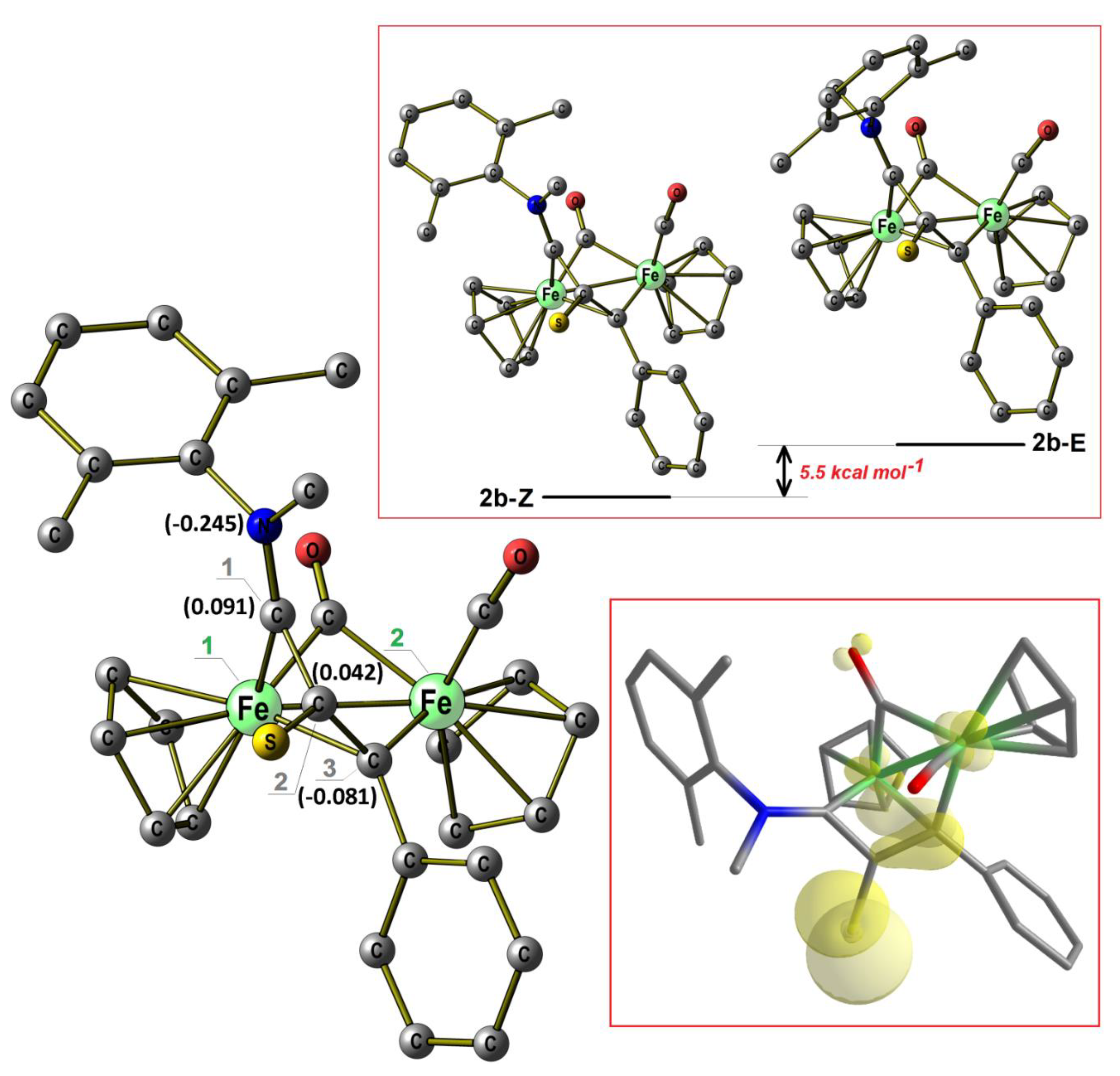
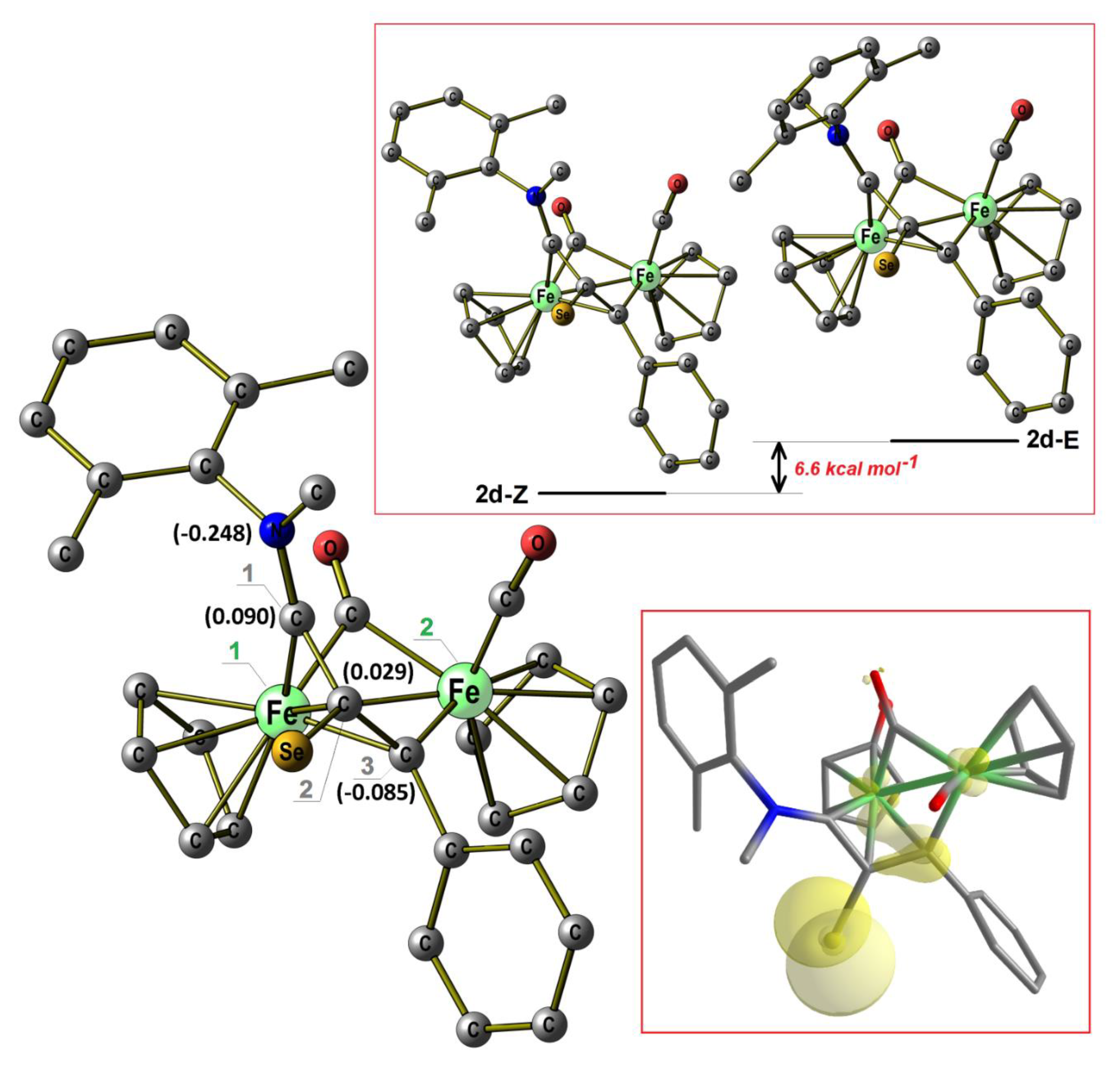
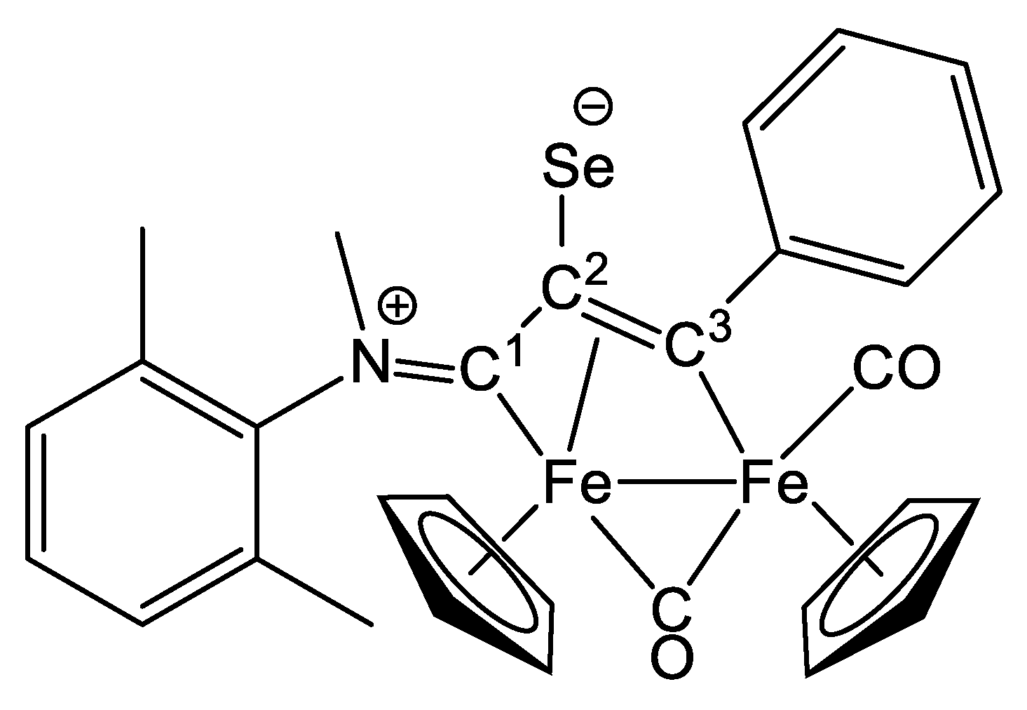
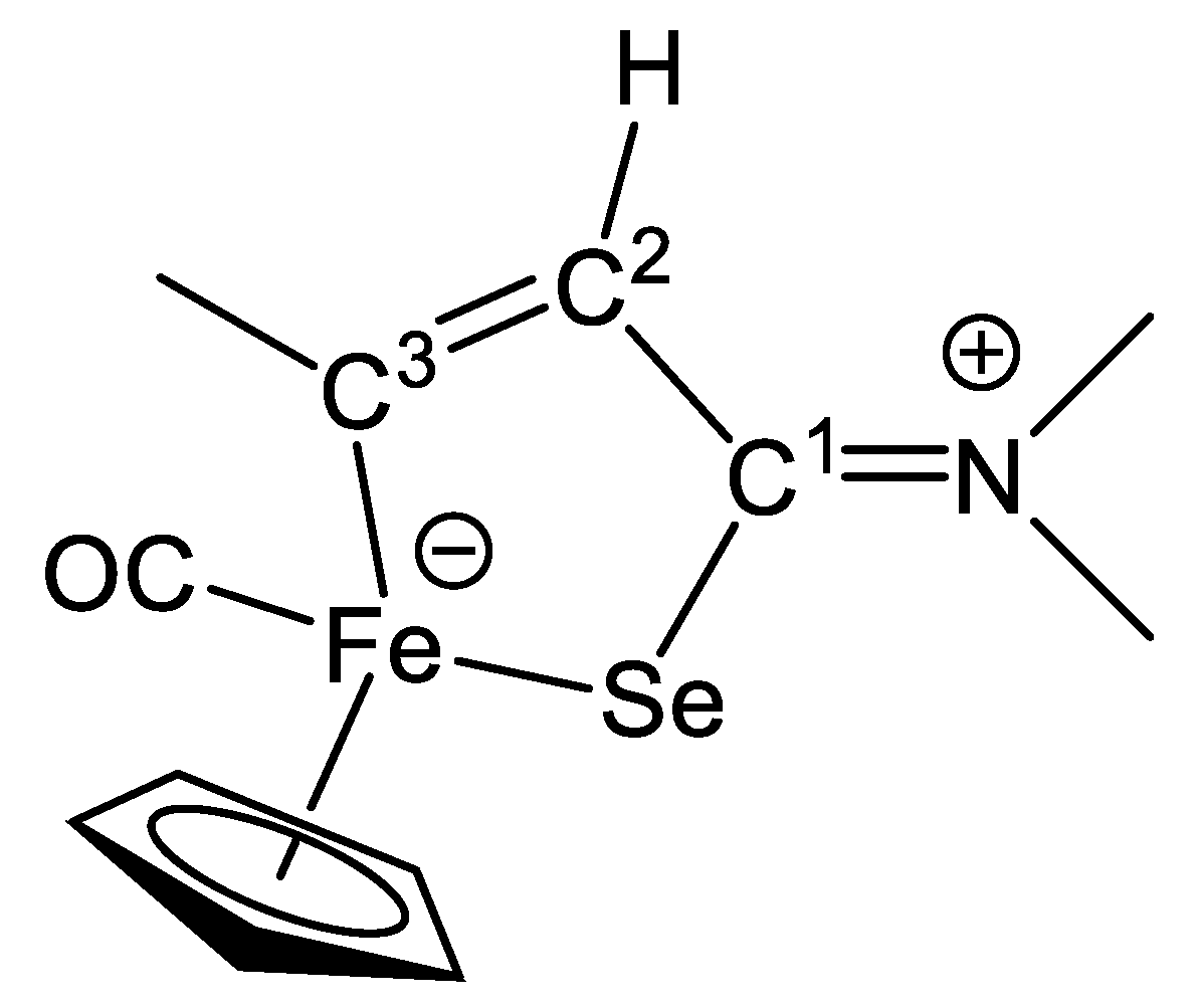
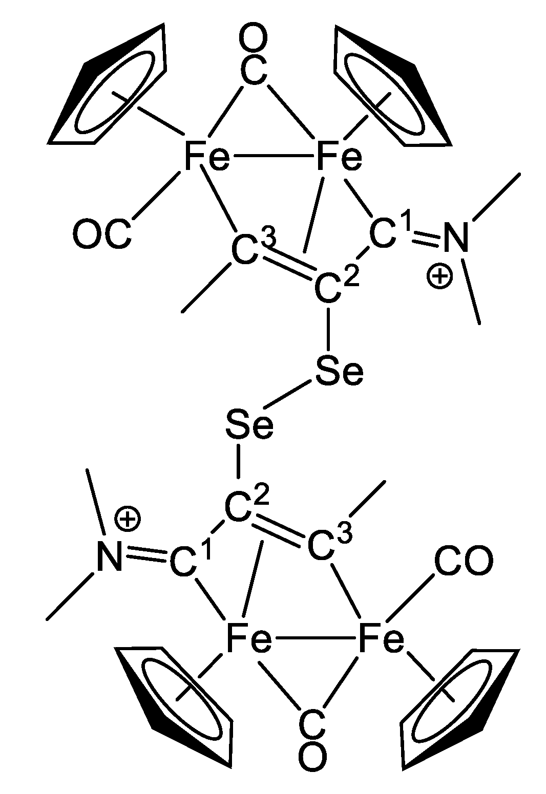
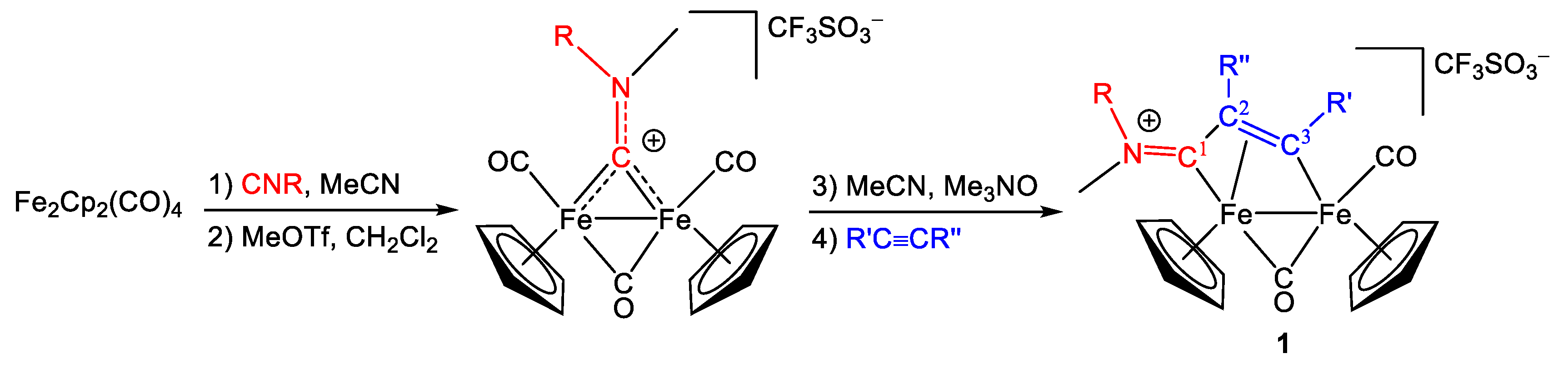
2.1. Synthesis and Characterization of Compounds, and DFT Analysis

Synthesis and Characterization of Compounds

Compounds 2a and 2c [48], 4c [49] and 6 [50] were previously reported, whereas 2b, 2d, 3, 4a, 4b, 4d, 5a, 5b, and 6 are novel (Scheme 2). Once isolated, 2ad slowly decompose in contact with air, whereas 36 resulted indefinitely air-stable. The sodium hydride(methoxide)-promoted dehydrogenative chalcogenylation of 1ac, as described previously [48], provides access to the zwitterionic complexes 2ad, in 60%-80% yields. This formal [C2H]+/C2E substitution (E = S, Se) presumably proceeds through the initial single-electron reduction of the cationic part of 1ac. Consistent with this hypothesis, the monoiron complex 3, maintaining the C2-H unit, is a side product of the reaction leading to 2a, and may be viewed as the result of selenium incorporation along a fragmentation process initiated by electron transfer to 1a [51]. The chalcogenido moiety in 2ad is readily oxidized with I2 to the dimeric iodide salts 4ad, containing an E-E bridge (77%–93%) [48]. Electrophilic methylation of 2ad affords 5ab (76%-86%). Instead, 6 is directly derived from 1a (80%), trapping the [SPh] fragment along the reaction of 1a with NaH [49].
According to combined X-ray diffraction analysis and NMR spectroscopy studies, the previously reported 2c and related R = Xyl containing complexes exist both in solution and in the solid state in the Z form, i.e., displaying the bulky xylyl group far from the chalcogen atom [48]. The salient NMR spectroscopic features of the new compounds, 2b and 2d, are in good agreement with those of 2c and analogues, thus indicating a Z configuration. For instance, the Cp rings and the methyl groups in the respective 1H NMR spectra are observed as follows: in 2b, at 4.59, 4.58 (Cp), 3.70 (Me) and 2.65, 2.16 ppm (Xyl); in 2d, at 4.62, 4.58 (Cp), 3.70 (Me) and 2.73, 2.16 ppm (Xyl); in [Fe2Cp2(CO)(μ-CO){μ-η13-C(4-C6H4Me)C(S)CN(Me)(Xyl)}] [47], at 4.59, 4.55 (Cp), 3.69 (Me) and 2.65, 2.16 ppm (Xyl). The latter complex differs from 2b in the presence of a 4-tolyl substituent in the place of Ph, and its structure was confirmed by X-ray diffraction.
DFT calculations confirm that the Z isomers of 2b and 2d are more stable than the E form by about 6 kcal mol−1 (Figure 1 and Figure 2). A comparison of computed bond lengths and angles indicates only small changes on replacing sulphur (2b) with selenium (2d). The most affected distance is Fe(2)-C(2), being 2.143 Å in 2b and 2.119 Å in 2d (CPCM/ωB97X calculations). The similarity between 2b and 2d is confirmed by the Mulliken population analysis, providing close values of partial charge for the μ-vinyliminium ligand in the two compounds. The higher stability of the Z isomers can be explained on the basis of the lower electrostatic repulsion between the chalcogen atom and the xylyl ring, as observable for instance in Figure S1 (Supporting information), where the electrostatic potential surfaces of E-2b and Z-2b are compared.
The structure of 3 was ascertained by single crystal X-ray diffraction (Figure 3, Table 1). Both C(1)-N(1) [1.28(6) and 1.27(6) Å for the two independent molecules present within the unit cell] and C(2)-C(3) [1.39(6) and 1.39(6) Å] distances show a significant double bond character, whereas C(1)-C(2) [1.46(6) and 1.43(6) Å] is essentially a C(sp2)-C(sp2) single bond with limited π-character. The Fe(1)-C(3) bond [1.96(4) and 1.92(5) Å] is elongated with respect to a pure terminal FeII-alkylidene, revealing a vinyl character [52,53,54,55]. The Fe(1)-Se(1) distance [2.391(8) and 2.357(9) Å] is in keeping with previously reported iron(II)-selenide bonds [56,57,58,59,60]. The perfectly planar five-membered ring [mean deviation from the Fe(1)-Se(1)-C(1)-C(2)-C(3) least-squares plane 0.0094 and 0.0409 Å] can be described as a zwitterionic ferra-selenophene-iminium, and to the best of our knowledge is unprecedented in organometallic chemistry.
The 1H and 13C NMR spectra of 3 (acetone-d6 solution) display two resonances for the N-bound methyls, in accordance with the iminium description of the [C1-NMe2] moiety. Signals attributable to the C1, C2 and C3 carbons are observed at 218.6, 137.4 and 199.3 ppm, respectively, while the selenium centre is observed at 285.7 ppm in the 77Se NMR spectrum.
In both 2b and 2d, the HOMO is localized on a p-type orbital of E (E = S, Se) and to a lesser extent on C3, C2 and on the iron centers (see Figure 1 and Figure 2): this explains why the chalcogen atom E is the most probable site for molecular oxidation or electrophilic attack. The HOMO of 2d is located 0.21 eV higher than in 2b, so I2-oxidation of 2d to 4d is 6.5 kcal mol−1 more favorable than the analogous reaction for 2b. Presumably, 4b and 4d containing xylyl groups, maintain the Z configuration of the N-substituents adopted in their precursors 2b and 2d [48]. Indeed, the NMR spectra of 4ad suggest the presence of a single species in solution. On going from 2d to 4d, the Se center undergoes significant deshielding in the 77Se NMR spectrum (from 282.5 to 556.4 ppm). DFT-optimized structures of 4b and 4d are shown in Figures S2 and S3.
The salient IR and NMR features of 5ab are typical of cationic vinyliminium complexes. In particular, the NMR spectra of 5b closely resemble those reported for Z-[Fe2Cp2(CO)(μ-CO){μ-η13-C(4-C6H4Me)C(SH)CN(Me)(Xyl)}]CF3SO3, 5c, whose structure was confirmed by X-ray diffraction [e.g.,: δ(1H, 5b/5c) = 5.16/4.99, 5.10/4.97 (Cp), 3.54/3.54 (NMe), 2.61/2.58, 2.07/2.04 (Xyl) ppm; δ(13C, 5b/5c)/ppm = 227.9/227.8 (C1); 68.4/68.2 (C2); 205.6/208.5 (C3)] [39]. In the 77Se NMR spectrum of 5a (acetone-d6 solution), the selenido unit gives rise to a signal at 187.4 ppm.

2.2. Electrochemistry

Compounds 4c and 5a were selected for electrochemical characterization in acetonitrile, which was extended to the respective precursors 1c and 2c, and also to 1a. The main results are summarized in Table 2, and all cyclic voltammetric profiles (referred to the ferrocenium/ferrocene redox couple) are provided in the Supporting Information (Figures S4–S14). In general, the investigated complexes exhibit one electrochemical reduction process, which occurs reversibly for 1a and 5a on the time scale of the experiment, respectively at −1.37 V and −1.29 V. However, the reduction observed for 1a is complicated by either two different processes occurring at very similar potentials, or a single process occurring in two steps (Figure S6). As a consequence, a slightly high peak-to-peak separation (∆Ep) of 108 mV was recognized. Furthermore, 1a displays an irreversible oxidation at +0.65 V, whereas in the case of 5a several irreversible oxidation reactions were detected in the potential range from −0.44 V to +0.66 V.
As discussed above, the chalcogenido moiety of 2c can be chemically oxidized to 4c (Scheme 2), and the same conversion was investigated using electrochemical techniques. As expected, the cyclic voltammogram (CV) of 2c shows an irreversible oxidation at +0.12 V, ascribable to the generation of the cationic part of 4c. Correspondingly, the CV profile of 4c shows an irreversible reduction at −0.78 V, that could be assigned to the formation of 2c [61]. Further considerations are prevented due to the presence of iodide as the counteranion in 4c, which is redox active and leads to the deposition of degradation products on the surface of the working electrode.

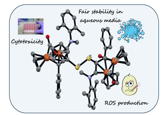
2.3. Cytotoxicity Studies and Stability in Aqueous Media

The air sensitive compounds 2ad were excluded from the biological tests. The remaining compounds were preliminarily evaluated for their stability in aqueous media (data summarized in Table 6). The ionic compounds 4ad, 5ab and 6, which are slightly soluble in water, and 3 did not undergo significant modification in DMSO-d6/D2O solution after 72 h at ca. 37 °C, according to 1H NMR spectroscopy (see Experimental for details). Approximately 50% degradation of 4b to unidentified species was detected after a further 72 h following addition of NaCl to the solution, whereas 4a,c,d did not change under the same conditions. IR spectroscopy was used to estimate the stability of 4ad, 5a and 6 in contact with cell culture medium at 37 °C. Compounds 4b, 4d, 5a, and 6 remained intact after 72 h, whereas 4a and 4c were recovered at the end of the experiment together with other carbonyl species. Indeed, a significant amount of 2c was detected to be produced from 4c.
Compounds 36 were assessed for their cytotoxicity against cisplatin sensitive (A2780) and cisplatin resistant (A2780cisR) human ovarian carcinoma cells, and the non-tumoral human embryonic kidney (HEK-293) cell line (see Table 3 and Experimental for details). Cisplatin and [(η6-p-cymene)RuCl2P-pta)] (RAPTA-C)[62] were evaluated as positive and negative controls, respectively.
Three tetrairon complexes, i.e., 4b4d, containing a S–S or a Se–Se bridge, and the diiron vinyliminium complexes 5b and 6, bearing a thioether function, possess potent cytotoxicity against the cancer cell lines, with IC50 values in the low micromolar/nanomolar range. In particular, the activity of 4b, 4d, and 5d is superior than that of cisplatin and appears to overcome resistance issues, since comparable IC50 values were determined on the A2780 and A2780cisR cell lines. However, selectivity is not observed compared to the HEK-293 cell line, apart from a moderate selectivity shown by 5a. The introduction of a Se–Se bridge on 1a leads to a dramatic decrease in activity, the diselenide derivative 4a being inactive towards all the investigated cell lines. In general, the strongest cytotoxic effect promoted by 4b,d, compared to 4a,c, reflects the higher stability in aqueous media of the former respect to the letter (see above).

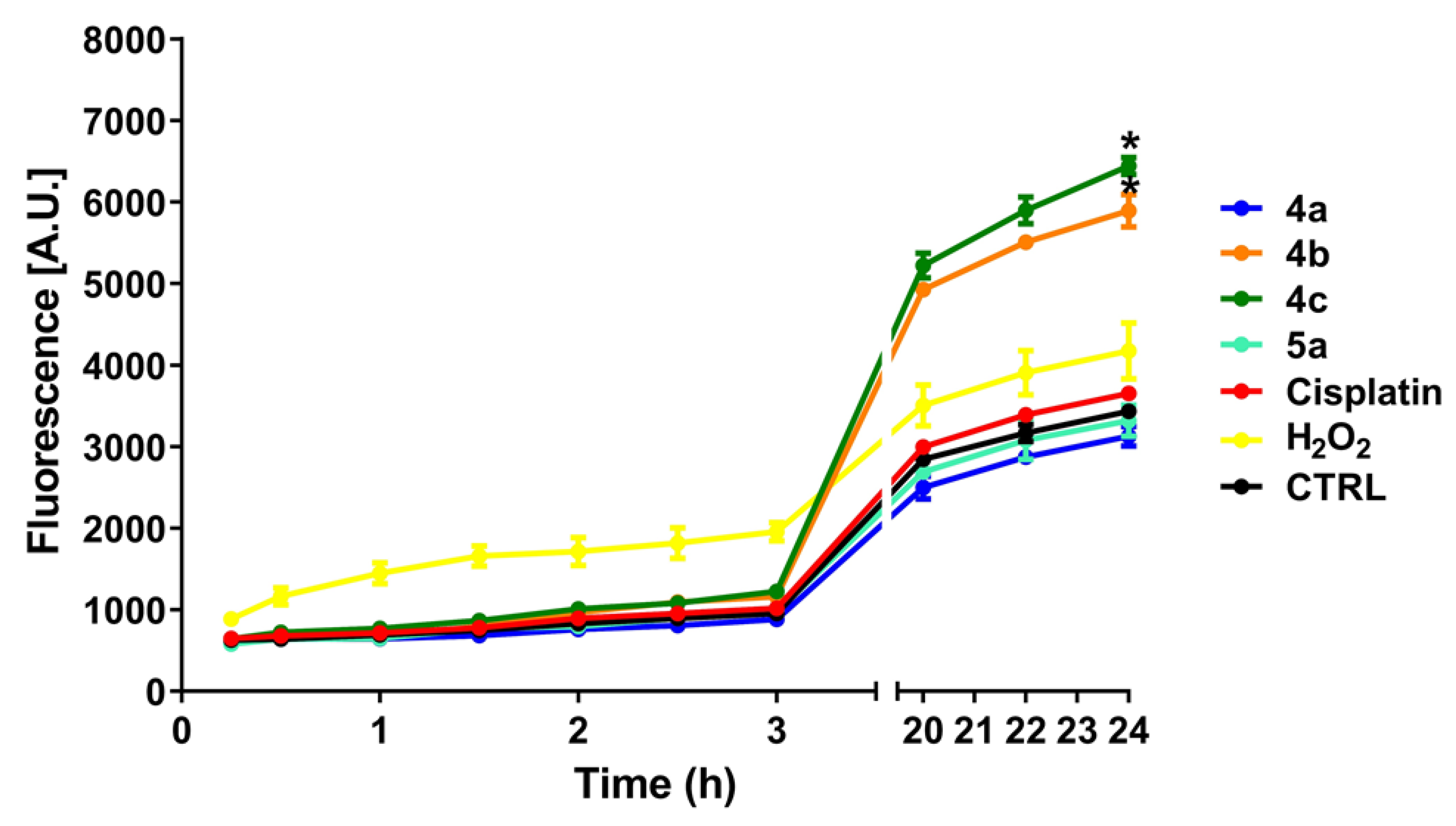
2.4. ROS Production and NADH Oxidation

We previously hypothesized that the cytotoxicity of diiron vinyliminium compounds, 1, is mainly ascribable to redox mechanisms (see Introduction). As suggested by the DFT outcomes, electrochemical investigations and stability data (see above), the tetrairon-bis-cationic complexes 4ad are susceptible to relatively facile reduction due to feasible disulphide(diselenide) to sulphide(selenide) conversion. Even the reduction of the selenido-vinyliminium 5a appears slightly more favorable with respect to analogous non-functionalized vinyliminium complexes (Table 2) [63]. Therefore, the cytotoxicity of the S- and Se-derivatives, and especially 4bd, is expected to involve interference of cellular redox processes. In order to investigate this aspect, we assessed the production of intracellular ROS levels induced by a selection of complexes (fluorescence measurements, using the peroxide-sensitive probe DCFH-DA). Thus, A2780 cells were continuously exposed to 4a, 4b, 4c, 5a, cisplatin (as a reference compound) and H2O2 (as a positive control). Treatment with 4b and 4c showed a significant increase in the level of ROS after ca. 20 h of treatment with respect to the positive control (Figure 4). Instead, 4a and 5a stimulated a ROS production close to that recorded for the basal levels; moreover, 4a did not show a significant effect even at higher concentration (100 µM). The marked difference in behavior between 4a (non cytotoxic) and 4bd indicates that the stimulation of ROS production could be indeed a privileged way of antiproliferative action for 4bd.
In order to further evaluate the ability of compounds to interfere with physiological redox processes, we determined the catalytic activity of 4a, 4c, 4d, 5a, and 6 in the aerobic oxidation of NADH, using a previously documented UV-Vis method (Table 4) [64]. Indeed, nicotinamide adenine dinucleotide (NAD+) and its reduced form (NADH) are important cofactors contributing to the maintenance of redox balance in cells [65], and the alteration of the NADH/NAD+ ratio has been implicated in the anticancer activity of various late transition metal complexes [64,66]. Cationic diiron vinyliminium compounds without chalcogen-functions, i.e., 1a and 1c, were also included in this study for comparison, together with FeSO4 as a reference compound. All tetrairon compounds 4a, 4c, 4d displayed a moderate catalytic activity on NADH oxidation, comparable (or slightly superior) to that of their diiron precursors (1a, 1c). Surprisingly, the diiron compounds 5a and 6, featuring selenoether/thioether moieties, were able to retard the oxidation of NADH with respect to the blank experiment. Notably, TONs at 25 h were significantly lower for 5a and 6 than for FeSO4, the latter exhibiting no catalytic activity.

3. Experimental

3.1. Synthetic Procedures and Compound Characterization

General details. The preparation, purification and isolation of compounds were carried out under a N2 atmosphere using standard Schlenk techniques; once obtained, 36 were stored in air and 2ad were stored under N2. Solvents were purchased from Merck and distilled before use under N2 from appropriate drying agents. Organic reactants (TCI Europe or Merck) were commercial products of the highest purity available. Compounds 1a-e [39,42], 2a,c [48], 4c [49], and 6 [50] were prepared according to published procedures. Chromatography separations were carried out on columns of deactivated alumina (Merck, 4% w/w water). Infrared spectra of solutions were recorded on a Perkin Elmer Spectrum 100 FT-IR spectrometer with a CaF2 liquid transmission cell (2300–1500 cm−1 range); IR spectra were processed with Spectragryph software [67]. NMR spectra were recorded at 298 K on a Bruker Avance II DRX400 instrument equipped with a BBFO broadband probe. Chemical shifts (expressed in parts per million) are referenced to the residual solvent peaks (1H, 13C) [68], or to external standard (77Se, SeMe2). 1H and 13C NMR spectra were assigned with the assistance of 1H-13C (gs-HSQC and gs-HMBC) correlation experiments [69]. Elemental analyses were performed on a Vario MICRO cube instrument (Elementar).
Synthesis of [Fe2Cp2(CO)(μ-CO){μ-η13-C3(Ph)C2(E)C1N(Me)(Xyl)}] (E = S, 2b; E = Se, 2d).
Compound 2b was prepared using the procedure reported in the literature for 2a and 2c [48], and a slight modification of the procedure was employed for 2d.
[Fe2Cp2(CO)(μ-CO){μ-η13-C3(Ph)C2(S)C1N(Me)(Xyl)}],2b(Chart 1).
From 1b (0.70 mmol), S8 (ca. 10 eq.) and NaH (4 eq.), see ref. [48]. Dark-green solid, yield 60%. Eluent for chromatography: CH2Cl2. Anal. calcd. for C30H27Fe2NO2S: C, 62.41; H, 4.71; N, 2.43; S, 5.56. Found: C, 63.06; H, 4.80; N, 2.40; S, 5.40. IR (CH2Cl2): ῦ/cm−1 = 1964vs (CO), 1791s (μ-CO), 1600m (C1N), 1581w (arom C-C). 1H NMR (CDCl3): δ/ppm = 7.68–7.28 (m, 8 H, C6H5 + C6H3Me2); 4.59, 4.58 (s, 10 H, Cp); 3.70 (s, 3 H, NMe); 2.65, 2.16 (s, 6 H, C6H3Me2). 13C{1H} NMR (CDCl3): δ/ppm = 264.2 (μ-CO); 235.4 (C1); 212.8 (CO); 195.7 (C3); 156.8 (ipso-C6H5); 142.3 (ipso-C6H3Me2); 135.7, 134.9, 129.3, 129.0, 127.8, 126.4 (C6H5 + C6H3Me2); 113.0 (C2); 90.6, 89.4 (Cp); 45.9 (NMe); 18.5, 18.0 (C6H3Me2). C2 observed via g-HSQC experiment.
[Fe2Cp2(CO)(μ-CO){μ-η13-C3(Ph)C2(Se)C1N(Me)(Xyl)}], 2d(Chart 2).
A solution of 1b (180 mg, 0.259 mmol) in THF (15 mL) was treated with gray Se (200 mg, 2.53 mmol) followed by NaOMe (35 mg, 0.648 mmol). The mixture was allowed to stir at room temperature for 1 h, then it was filtered through a short alumina pad, using neat THF as eluent, under protection from air. The filtrate was dried under vacuum. The resulting residue was dissolved in CH2Cl2 and the solution was charged on alumina. Elution with CH2Cl2 removed the impurities and a green band was collected using THF as eluent. The title compound was isolated as a brown solid upon removal of the solvent under vacuum. Yield 129 mg, 80%. Anal. calcd. for C30H27Fe2NO2Se: C, 57.73; H, 4.36; N, 2.24. Found: C, 57.61; H, 4.44; N, 2.18. IR (CH2Cl2): ῦ/cm−1 = 1967vs (CO), 1794s (μ-CO), 1604w (C1N), 1583w (arom C-C). 1H NMR (CDCl3): δ/ppm = 7.69–7.25 (m, 8 H, Ph + C6H3Me2); 4.62, 4.58 (s, 10 H, Cp); 3.70 (s, 3 H, NMe); 2.73, 2.16 (s, 6 H, C6H3Me2). 13C{1H} NMR (CDCl3): δ/ppm = 262.8 (μ-CO); 229.7 (C1); 212.4 (CO); 198.6 (C3); 157.7 (ipso-Ph); 141.9 (ipso-C6H3); 136.1, 135.0, 129.6, 129.1, 129.0, 128.8, 128.2, 128.0, 125.9, 125.3 (Ph + C6H3Me2); 90.7, 90.4 (Cp); 89.6 (C2); 47.0 (NMe); 18.6, 18.3 (C6H3Me2). 77Se NMR (CDCl3): δ/ppm = 282.5.
Synthesis of [FeCp(CO){SeC1(NMe2)C2HC3(Me)}], 3(Chart 3).
The reaction mixture for the synthesis of 2a was obtained as described in the literature, from 1a, gray selenium and NaH [48]. This mixture was filtered through a short alumina pad using THF as eluent, then the volatiles were removed under vacuum. Subsequent alumina chromatography of the residue led to isolate a red fraction using neat diethyl ether as eluent, corresponding to 3. Compound 3 was isolated as an air stable, red solid upon removal of the solvent under vacuum. Yield 16%. Anal. calcd. for C12H15FeNOSe: C, 44.48; H, 4.67; N, 4.32. Found: C, 44.12; H, 4.51; N, 4.39. IR (CH2Cl2): ῦ/cm−1 = 1921vs (CO), 1530m (C3=C2). 1H NMR (acetone-d6): δ/ppm = 7.36 (s, 1 H, C2-H); 4.59 (s, 5 H, Cp); 3.45, 3.28 (s, 6 H, NMe2); 2.77 (s, 3 H, C3-Me). 13C{1H} NMR (acetone-d6): δ/ppm = 252.3 (CO); 218.6 (C1); 199.3 (C3); 137.4 (C2); 82.3 (Cp); 44.8, 42.5 (NMe2); 40.1 (C3Me). 77Se NMR (acetone-d6): δ/ppm = 285.7. Crystals suitable for X-ray analysis were obtained from a diethyl ether solution layered with pentane and stored at −30 °C.
Synthesis of [Fe2Cp2(CO)(μ-CO){μ-η13-C3(R’)C2(E)C1N(Me)(R)}]2[I]2 (R = R’ = Me, E = Se,4a; R = Xyl, R’ = Ph, E = S,4b; R = Xyl, R’ = Ph, E = Se,4d).
The title products were prepared using the procedure reported in the literature for 4c [49].
[Fe2Cp2(CO)(μ-CO){μ-η13-C3(Me)C2(Se)C1NMe2}]2[I]2, 4a (Chart 4).
From 2a and I2, see ref. [49]. Red solid, yield 84%. Anal. calcd. for C36H38Fe4I2N2O4Se2: C, 36.10; H, 3.20; N, 2.34. Found: C, 36.05; H, 3.26; N, 2.41. IR (CH2Cl2): ῦ/cm−1 = 1995vs (CO), 1815s (μ-CO), 1671m (C2C1N). 1H NMR (acetone-d6): δ/ppm = 5.75, 5.34 (s, 20 H, Cp); 4.24, 4.12 (s, 12 H, NMe + C3Me); 3.62 (s, 6 H, NMe2). 13C{1H} NMR (acetone-d6): δ/ppm = 252.9 (μ-CO); 220.9 (C1); 210.0 (CO); 208.7 (C3); 92.1, 89.1 (Cp); 57.1 (C2); 51.6, 45.9 (NMe); 41.1 (C3Me). 77Se NMR (DMSO-d6): δ/ppm = 519.8.
[Fe2Cp2(CO)(μ-CO){μ-η13-C3(Ph)C2(S)C1N(Me)(Xyl)}]2[I]2, 4b(Chart 5).
From 2b and I2, see ref. [49]. Dark-red solid, yield 77%. Anal. calcd. for C60H54Fe4I2N2O4S2: C, 51.17; H, 3.86; N, 1.99; S, 4.55. Found: C, 51.02; H, 3.94; N, 2.06; 4.69. IR (CH2Cl2): ῦ/cm−1 = 1994vs (CO), 1830s (μ-CO), 1611m (C2C1N), 1586w (arom C-C). 1H NMR (CD2Cl2): δ/ppm = 7.81–7.29 (m, 8 H, C6H5 + C6H3Me2); 5.10, 5.08 (s, 10 H, Cp); 3.21 (s, 3 H, NMe); 2.66, 2.10 (s, 6 H, C6H3Me2). 13C{1H} NMR (CD2Cl2): δ/ppm = 249.9 (μ-CO); 227.3 (C1); 210.1, 209.2 (CO + C3); 152.7 (ipso-C6H5); 140.1 (ipso-C6H3Me2); 134.3-126.7 (C6H5 + C6H3Me2); 90.6, 89.4 (Cp); 65.8 (C2); 45.9 (NMe); 18.8, 18.1 (C6H3Me2). NMe overlapped with solvent signal.
[Fe2Cp2(CO)(μ-CO){μ-η13-C3(Ph)C2(Se)C1N(Me)(Xyl)}]2[I]2, 4d(Chart 6).
From 2b and I2, see ref. [49]. Brown solid, yield 93%. Anal. calcd. for C60H54Fe4I2N2O4Se2: C, 47.97; H, 3.62; N, 1.86. Found: C, 47.85; H, 3.68; N, 1.93. IR (CH2Cl2): ῦ/cm−1 = 1994vs (CO), 1825s (μ-CO), 1616m (C2C1N), 1586w (arom C-C). 1H NMR (CD3CN): δ/ppm = 7.85–7.45, 7.36, 7.01 (m, 8 H, C6H5 + C6H3Me2); 5.04, 4.98 (s, 10 H, Cp); 3.49 (s, 3 H, NMe); 2.52, 2.14 (s, 6 H, C6H3Me2). 77Se NMR (DMSO-d6): δ/ppm = 556.4.
Synthesis of [Fe2Cp2(CO)(μ-CO){μ-η13-C3(R’)C2(EMe)C1N(Me)(R)}]I (R = R’ = Me, E = Se,5a; R = Xyl, R’ = Ph, E = S,5b).
General procedure. Compound 2ab (ca. 0.50 mmol) was dissolved in CH2Cl2 (15 mL), and MeI (1.5 equivalents) was added to the solution. The resulting mixture was stirred at room temperature for 2 h, and then charged on an alumina column. Elution with CH2Cl2 allowed to separate impurities, then the fraction corresponding to the product was collected using MeCN/MeOH (95/5 v/v) as eluent.
[Fe2Cp2(CO)(μ-CO){μ-η13-C3(Me)C2(SeMe)C1NMe2)}]I, 5a(Chart 7).
From 2a and MeI. Brown solid, yield 86%. Eluent for chromatography: MeOH. Anal. calcd. for C27H31Fe2INO2Se: C, 45.10; H, 4.35; N, 1.95. Found: C, 44.90; H, 4.27; N, 1.98. IR (CH2Cl2): ῦ/cm−1 = 1992vs (CO), 1813s (μ-CO), 1667m (C2C1N). 1H NMR (acetone-d6): δ/ppm = 5.59, 5.24 (s, 10 H, Cp); 4.13 (s, 3 H, C3Me); 4.03, 3.32 (s, 6 H, NMe2); 2.35 (s, 3 H, SeMe). 13C{1H} NMR (acetone-d6): δ/ppm = 255.4 (μ-CO); 220.3 (C1); 210.3 (CO); 205.0 (C3); 91.3, 88.9 (Cp); 65.1 (C2); 47.6, 44.9 (NMe); 39.1 (C3Me); 6.7 (SeMe). 77Se NMR (DMSO-d6): δ/ppm = 187.4.
[Fe2Cp2(CO)(μ-CO){μ-η13-C3(Ph)C2(SMe)C1N(Me)(Xyl)}]I, 5b(Chart 8).
From 2b and MeI. Dark-brown solid, yield 76%. Eluent for chromatography: THF. Anal. calcd. for C31H30Fe2INO2S: C, 51.77; H, 4.20; N, 1.95; S, 4.46. Found: C, 51.65; H, 4.26; N, 2.03; S, 4.40. IR (CH2Cl2): ῦ/cm−1 = 1993vs (CO), 1829s (μ-CO), 1611m (C2C1N), 1587w (arom C-C). 1H NMR (CDCl3): δ/ppm = 7.91, 7.75, 7.54–7.41, 6.98 (m, 8 H, C6H5 + C6H3Me2); 5.16, 5.10 (s, 10 H, Cp); 3.54 (s, 3 H, NMe); 2.61, 2.07 (s, 6 H, C6H3Me2); 2.12 (s, 3 H, SMe). 13C{1H} NMR (CDCl3): δ/ppm = 250.7 (μ-CO); 227.9 (C1); 210.7 (CO); 205.6 (C3); 152.5 (ipso-C6H5); 140.5 (ipso-C6H3Me2); 135.8, 134.2, 134.1, 130.2, 130.0, 129.2, 128.1, 127.4, 127.3, 127.1, 125.5 (C6H5 + C6H3Me2); 93.3, 88.8 (Cp); 68.4 (C2); 51.5 (NMe); 19.6 (SMe); 18.2, 18.0 (C6H3Me2).

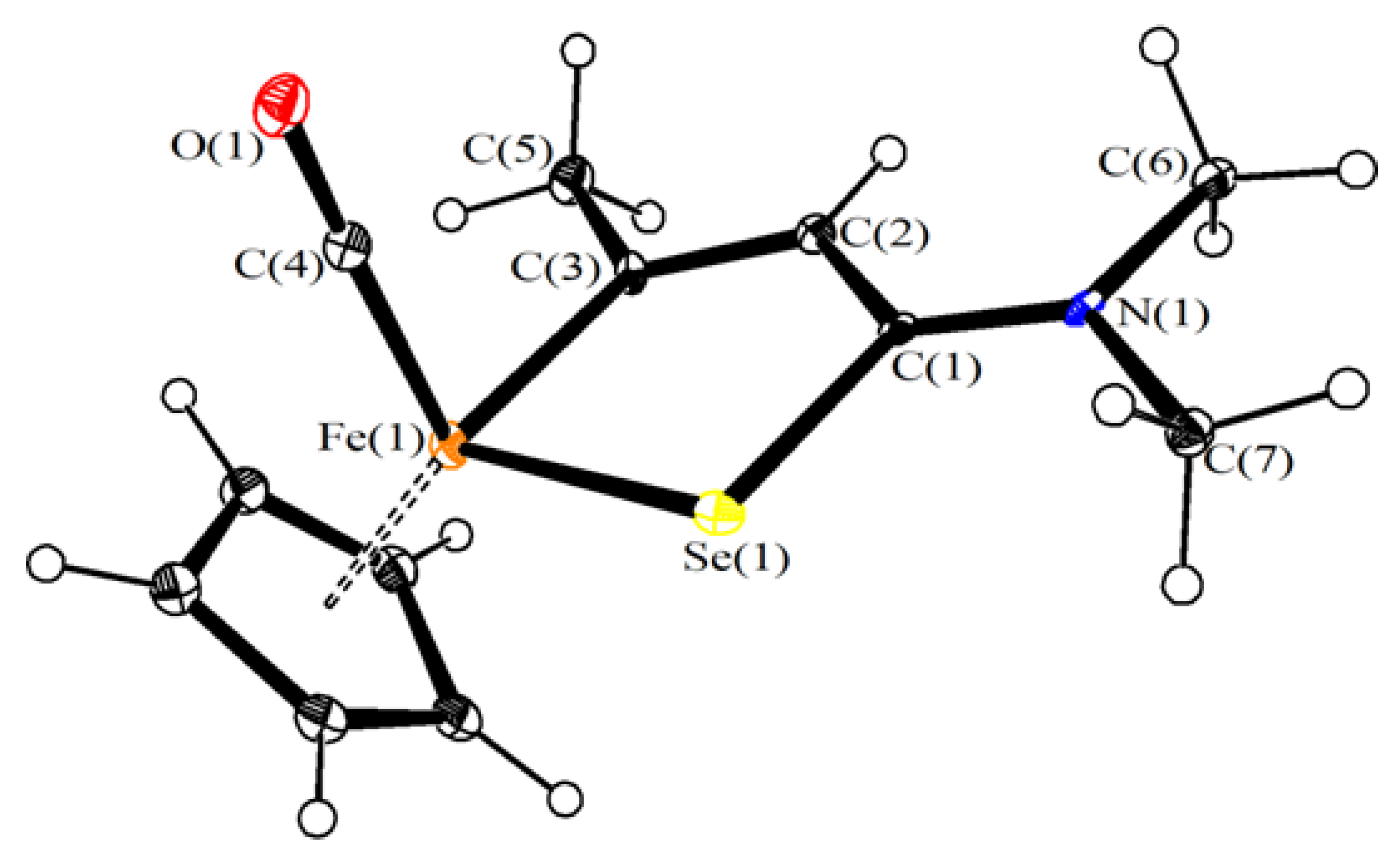
3.2. X-Ray Crystallography

Crystal data and collection details for 3 are reported in Table 5. Data were recorded on a Bruker APEX II diffractometer equipped with a PHOTON100 detector using Mo–Kα radiation. The crystal appeared to be non-merohedrally twinned. The program CELL_NOW (G. M. Sheldrick, CELL_NOW, Version 2008/4, 2008) was used in order to determine the two twin domains and their orientation matrices. After integration, data were corrected for Lorentz polarization and absorption effects (empirical absorption correction TWINABS) [70]. The structure was solved by direct methods and refined by full-matrix least-squares based on all data using F2 [71]. Hydrogen atoms were fixed at calculated positions and refined using a riding model. All non-hydrogen atoms were refined with anisotropic displacement parameters. Refinement was performed using the instruction HKLF 5 in SHELXL and one BASF parameter, which refined as 0.276(7). Because of the twinning, several restraints were applied during refinement. All the atoms were restrained to have similar thermal parameters (SIMU line in SHLEXL, s.u. 0.01). All C, O, and N atoms were restrained to isotropic like behavior (ISOR line in SHELXL, s.u. 0.01).

3.3. Computational Studies

The electronic structures of the compounds were optimized using the range-separated ωB97X DFT functional [72,73,74] in combination with Ahlrichs’ split-valence-polarized basis set [75]. The C-PCM implicit solvation model was added to ωB97X calculations, considering chloroform as a continuous medium [76,77].
Preliminary optimizations were carried out using the hybrid-GGA EDF2 functional [78] in combination with the 6-31G(d,p) basis set [79]. The stationary points were characterized by IR simulations (harmonic approximation), from which zero-point vibrational energies and thermal corrections (T = 25 °C) were obtained. Simulated IR spectra were used to assign the experimentally observed signals [80]. The software used were Gaussian 09 (Gaussian, Inc: Wallingford, CT, USA) [81] and Spartan ‘16 [82]. Cartesian coordinates of the DFT-optimized structures are collected in a separated .xyz file.

3.4. Stability in Aqueous Solutions

Each compound (ca. 10 mg; 3, 4ad, 5ab, 6) was dissolved in DMSO-d6 (0.4 mL), then the solution was diluted with variable volumes of D2O. The resulting solution was kept at 37 °C for 72 h. Subsequent 1H NMR spectra revealed the presence of the respective starting materials together with minor decomposition products (<10%). NaCl was added in ca. 0.05 M concentration to the solutions containing 4ad, and the obtained mixtures were kept at 37 °C for 72 h before 1H NMR spectra were recorded (Table 6). In order to perform tests in contact with a cell culture medium, compounds 4cd, 5a and 6 (ca. 4 mg) were dissolved in DMSO (ca. 1 mL) in a glass tube, then RPMI-1640 medium with sodium bicarbonate, without l-glutamine and phenol red (ca. 3 mL, Merck), was added. The resulting mixture was maintained at 37 °C for 72 h, then it was allowed to cool to room temperature. Dichloromethane (ca. 4 mL) was added, and the mixture was vigorously shaken. An aliquot of the organic phase was analyzed by IR spectroscopy (Table 6).

3.5. Electrochemistry

Cyclic voltammograms were measured under an atmosphere of argon using standard Schlenk techniques with a Palmsens4 potentiostat by working with anhydrous and degassed solutions of acetonitrile (MeCN). MeCN was dried and distilled under Ar from the appropriate drying agent (CaH2), and thoroughly deoxygenated with Ar prior to use. The samples were measured at a concentration of 0.1 M and using 0.1 M NBu4PF6 (Merck) as conductive salt. A glassy carbon electrode was used as working electrode, a coiled platinum wire as counter electrode, and a silver wire as a pseudo-reference electrode. Ferrocene (or decamethylferrocene) was added as an internal standard and all spectra were referenced to the ferrocenium/ferrocene couple (Fc+/Fc).

3.6. Cell Culture and Cytotoxicity Studies

Human ovarian carcinoma (A2780 and A2780cisR) cell lines were obtained from the European Collection of Cell Cultures. The human embryonic kidney (HEK-293) cell line was obtained from ATCC (Merck, Buchs, Switzerland). Penicillin streptomycin, RPMI 1640 GlutaMAX (where RPMI = Roswell Park Memorial Institute), and DMEM GlutaMAX media (where DMEM = Dulbecco’s modified Eagle medium) were obtained from Life Technologies, and fetal bovine serum (FBS) was obtained from Merck. The cells were cultured in RPMI 1640 GlutaMAX (A2780 and A2780cisR) and DMEM GlutaMAX (HEK-293) media containing 10% heat-inactivated FBS and 1% penicillin streptomycin at 37 °C and CO2 (5%). The A2780cisR cell line was routinely treated with cisplatin (2 μM) in the media to maintain cisplatin resistance. The cytotoxicity was determined using the 3-(4,5-dimethyl 2-thiazolyl)-2,5-diphenyl-2H-tetrazolium bromide (MTT) assay [83]. Cells were seeded in flat-bottomed 96-well plates as a suspension in a prepared medium (100 μL aliquots and approximately 4300 cells/well) and preincubated for 24 h. Stock solutions of compounds were prepared in DMSO and were diluted in medium. The solutions were sequentially diluted to give a final DMSO concentration of 0.5% and a final compound concentration range (0–200 μM). Cisplatin and RAPTA-C [62] were tested in aqueous solution as a positive (0–100 μM) and negative (200 μM) controls, respectively. The compounds were added to the preincubated 96-well plates in 100 μL aliquots, and the plates were incubated for a further 72 h. MTT (20 μL, 5 mg/mL in Dulbecco’s phosphate buffered saline) was added to the cells, and the plates were incubated for a further 4 h. The culture medium was aspirated and the purple formazan crystals, formed by the mitochondrial dehydrogenase activity of vital cells, were dissolved in DMSO (100 μL/well). The absorbance of the resulting solutions, directly proportional to the number of surviving cells, was quantified at 590 nm using a SpectroMax M5e multimode microplate reader (using SoftMax Pro software, version 6.2.2). The percentage of surviving cells was calculated from the absorbance of wells corresponding to the untreated control cells. The reported IC50 values are based on the means from two independent experiments, each comprising four tests per concentration level.

3.7. ROS Production Assessment

The intracellular increase of reactive oxygen species (ROS) upon treatment with the analyzed complexes was measured by using the DCFH-DA (2′,7′-dichlorodihydrofluorescein diacetate, Merck) assay, based on cellular uptake of the non-fluorescent diacetate following deacetylation by esterases (2′,7′-dichlorodihydrofluorescein, DCFH) and oxidation to the fluorescent dichlorofluorescein (2′,7′-dichloro-fluorescein, DCF) [84]. A2780 cells were seeded at concentration of 4300 cells/well/90 µL of complete growth medium into 96-well plates and allowed to proliferate for 24 h. Then cells were treated following the manufacturer’s protocol. Briefly, the culture medium was supplemented with 100 µL of the fluorogenic probe solution and cells were incubated under standard tissue culture conditions of 5% CO2 at 37 °C. After 1 h, the cells were exposed with a final concentration of 10 µM of the tested compounds and maintained at 5% CO2 at 37 °C; H2O2 100 µM was used as a positive control. Stock solutions of compounds were prepared as described above. Cells incubated with DMSO at a concentration of 0.1% in supplemented RPMI were used as control. The fluorescence was measured over 24 h with an excitation wavelength of 485 nm and with a 535 nm emission filter by Multilabel Counter (PerkinElmer, Waltham, USA). Analysis was conducted in triplicate and experimental data were reported as mean ± SD. Statistical differences were analyzed using one-way analysis of variance (ANOVA) and a Tukey test was used for post hoc analysis. A p-value <0.05 was considered as statistically significant.

3.8. Catalytic NADH Oxidation

NADH was stored at –20 °C under N2; a stock NADH solution (2.3 × 10⁻⁴ mol L−1) was prepared in phosphate buffered aqueous solution (Na2HPO4/NaH2PO4; 5.5 × 10⁻³ mol∙L−1, pH = 7.2) and stored at 4 °C. Stock solutions of iron compounds (1a, 4a,c,d, 5a, 6; 2.0 × 10⁻⁴ mol∙L−1) were prepared in DMSO immediately before use. FeSO4 was used as a reference compounds (stock solution prepared in H2O). Solutions of each iron compound (0.35 mL) and NADH (6.6 mL) were mixed, resulting in a 5% DMSO aqueous solution containing 2.2 × 10⁻⁴ M NADH and 1.0∙× 10−5 M iron compound (4.5% mol). The solution was stirred at 37 °C for 25 h and periodically analyzed by UV-Vis spectroscopy (260–460 nm) using PMMA cuvettes (1.0 cm path-length). Turnover numbers were calculated as TON = c(0)/cFe∙[A(0) − A(t)]/A(0) where A is the absorbance at λmax = 339 nm; c(0) and cFe are the initial molar concentrations of NADH and the selected Fe compound, respectively (Table 4).

4. Conclusions

The bridging vinyliminium ligand in cationic diiron complexes can be modified by introducing sulphur- or selenium-functions according to well defined regio- and stereoselective reaction pathways. Some of the resulting, air stable diiron and tetrairon compounds display a strong antiproliferative activity against human ovarian carcinoma cell lines, the activity of some compounds on the A2780 cell line being superior than that of cisplatin and substantially maintained on the A2780cisR resistant cell line. Experiments suggest that the chalcogen function (especially the presence of an E–E bridge) is associated with good stability in aqueous media, enhancing interference of compounds with cellular redox processes.

Supplementary Materials

The following are available online: DFT structures, cyclic voltammograms, NMR spectra of products (signals around 0 ppm due to some silicon grease). CCDC reference number 1983327 (3) contains the supplementary crystallographic data for the X-ray studies reported in this paper. These data can be obtained free of charge at www.ccdc.cam.ac.uk/conts/retrieving.html (or from the Cambridge Crystallographic Data Centre, 12, Union Road, Cambridge CB2 1EZ, UK; fax: (internat.) +44-1223/336-033; e-mail: [email protected]).

Author Contributions

Conceptualization, F.M. and L.B.; methodology, G.A., L.K.B., L.B., S.S., E.F., S.B., S.Z., F.C.; software, M.B.; investigation, G.A., L.K.B., L.B., S.S., E.F., S.B., S.Z.; data curation, B.S., F.C.; writing—original draft preparation, F.M.; writing—review and editing, P.J.D., F.M., G.P.; supervision, P.J.D., F.M.; funding acquisition, G.P. All authors have read and agreed to the published version of the manuscript.

Funding

The work is supported by the University of Pisa (Fondi di Ateneo 2019).

Acknowledgments

We gratefully thank the University of Pisa (Fondi di Ateneo 2019) for financial support.

Conflicts of Interest

The authors declare no conflict of interest.

References

  1. Apps, M.G.; Choi, E.H.Y.; Wheate, N.J. The state-of-play and future of platinum drugs. Endocr. Relat. Cancer 2015, 22, R219–R233. [Google Scholar] [CrossRef] [PubMed] [Green Version]
  2. Gibson, D. The mechanism of action of platinum anticancer agents—What do we really know about it? Dalton Trans. 2009, 10681–10689. [Google Scholar] [CrossRef] [PubMed]
  3. Johnstone, T.C.; Suntharalingam, K.; Lippard, S.J. The Next Generation of Platinum Drugs: Targeted Pt(II) Agents, Nanoparticle Delivery, and Pt(IV) Prodrugs. Chem. Rev. 2016, 116, 3436–3486. [Google Scholar] [CrossRef] [PubMed] [Green Version]
  4. Raudenska, M.; Balvan, J.; Fojtu, M.; Gumulec, J.; Masarik, M. Unexpected therapeutic effects of cisplatin. Metallomics 2019, 11, 1182–1199. [Google Scholar] [CrossRef] [PubMed]
  5. Oun, R.; Moussa, Y.E.; Wheate, N.J. The side effects of platinum-based chemotherapy drugs: A review for chemists. Dalton Trans. 2018, 47, 6645–6653. [Google Scholar] [CrossRef] [PubMed]
  6. Min, Y.; Mao, C.Q.; Chen, S.; Ma, G.; Wang, J.; Liu, Y. Combating the Drug Resistance of Cisplatin Using a Platinum Prodrug Based Delivery System. Angew. Chem. 2012, 124, 6846–6851. [Google Scholar] [CrossRef]
  7. Siddik, Z.H. Cisplatin: Mode of cytotoxic action and molecular basis of resistance. Oncogene 2003, 22, 7265–7279. [Google Scholar] [CrossRef] [Green Version]
  8. Dasari, S.; Tchounwou, P.B. Cisplatin in cancer therapy: Molecular mechanisms of action. Eur. J. Pharmacol. 2014, 740, 364–378. [Google Scholar] [CrossRef] [Green Version]
  9. Kartalou, M.; Essigmann, J.M. Mechanisms of resistance to cisplatin. Mutat. Res. 2001, 478, 23–43. [Google Scholar] [CrossRef]
  10. Zhang, P.; Sadler, P.J. Advances in the design of organometallic anticancer complexes. J. Organomet. Chem. 2017, 839, 5–14. [Google Scholar] [CrossRef]
  11. Barry, N.P.E.; Sadler, P.J. Exploration of the medical periodic table: Towards new targets. Chem. Commun. 2013, 49, 5016–5041. [Google Scholar] [CrossRef] [PubMed] [Green Version]
  12. Štarha, P.; Trávníček, Z. Non-platinum complexes containing releasable biologically active ligands. Coord. Chem. Rev. 2019, 395, 130–145. [Google Scholar] [CrossRef]
  13. Santini, C.; Pellei, M.; Gandin, V.; Porchia, M.; Tisato, F.; Marzano, C. Advances in Copper Complexes as Anticancer Agents. Chem. Rev. 2014, 114, 815–862. [Google Scholar] [CrossRef] [PubMed]
  14. Alam, M.N.; Huq, F. Comprehensive review on tumour active palladium compounds and structure–activity relationships. Coord. Chem. Rev. 2016, 316, 36–67. [Google Scholar] [CrossRef]
  15. Nazarov, A.A.; Hartinger, C.G.; Dyson, P.J. Opening the lid on piano-stool complexes: An account of ruthenium(II)-arene complexes with medicinal applications. J. Organomet. Chem. 2014, 751, 251–260. [Google Scholar] [CrossRef]
  16. Rilak Simovica, A.; Masnikosa, R.; Bratsos, I.; Alessio, E. Chemistry and reactivity of ruthenium(II) complexes: DNA/protein binding mode and anticancer activity are related to the complex structure. Coord. Chem. Rev. 2019, 398, 113011. [Google Scholar] [CrossRef]
  17. Zou, T.; Lum, C.T.; Lok, C.N.; Zhang, J.J.; Che, C.M. Chemical biology of anticancer gold(iii) and gold(i) complexes. Chem. Soc. Rev. 2015, 44, 8786–8801. [Google Scholar] [CrossRef] [PubMed]
  18. Lazarevi, T.; Rilak, A.; Bugarcic, Z.D. Platinum, palladium, gold and ruthenium complexes as anticancer agents: Current clinical uses, cytotoxicity studies and future perspectives. Eur. J. Med. Chem. 2017, 142, 8–31. [Google Scholar] [CrossRef] [PubMed]
  19. Bratsos, T.; Gianferrara, E.; Alessio, C.G.; Hartinger, M.A.; Jakupec, B.K. Keppler, Bioinorganic Medicinal Chemistry; Alessio, E., Ed.; Wiley-VCH: Weinheim, Germany, 2011; pp. 151–174. [Google Scholar]
  20. Lord, R.; Zegke, M.; Henderson, I.R.; Pask, C.M.; Shepherd, H.J.; McGowan, P.C. β-Ketoiminato Iridium(III) organometallic complexes: Selective cytotoxicity towards colorectal cancer cells HCT116 p53-/-. Chem. Eur. J. 2019, 25, 495–500. [Google Scholar] [CrossRef] [PubMed]
  21. Fernandez-Gallardo, J.; Elie, B.T.; Sadhukha, T.; Prabha, S.; Sanau, M.; Rotenberg, S.A.; Ramos, J.W.; Contel, M. Heterometallic titanium–gold complexes inhibit renal cancer cells in vitro and in vivo. Chem. Sci. 2015, 6, 5269–5283. [Google Scholar] [CrossRef] [Green Version]
  22. Patra, M.; Gasser, G. The medicinal chemistry of ferrocene and its derivatives. Nat. Rev. Chem. 2017, 1, 1–12. [Google Scholar] [CrossRef]
  23. Braga, S.S.; Silva, A.M.S. A New Age for Iron: Antitumoral Ferrocenes. Organometallics 2013, 32, 5626–5639. [Google Scholar] [CrossRef]
  24. Jaouen, G.; Vessiéres, A.; Top, S. Ferrocifen type anti cancer drugs. Chem. Soc. Rev. 2015, 44, 8802–8817. [Google Scholar] [CrossRef] [PubMed] [Green Version]
  25. Agonigi, G.; Biancalana, L.; Lupo, M.G.; Montopoli, M.; Ferri, N.; Zacchini, S.; Binacchi, F.; Biver, T.; Campanella, B.; Pampaloni, G.; et al. Exploring the anticancer potential of diiron bis-cyclopentadienyl complexes with bridging hydrocarbyl ligands: Behavior in aqueous media and in vitro cytotoxicity. Organometallics 2020, 39, 645–657. [Google Scholar] [CrossRef]
  26. Hirschhäuser, C.; Velcicky, J.; Schlawe, D.; Hessler, E.; Majdalani, A.; Neudçrfl, J.M.; Prokop, A.; Wieder, T.; Schmalz, H.G. Nucleoside analogues with a 1,3-diene-Fe(CO)3 substructure: Stereoselective synthesis, configurational assignment, and apoptosis-inducing activity. Chem. Eur. J. 2013, 19, 13017–13029. [Google Scholar] [CrossRef]
  27. Cingolani, A.; Zanotti, V.; Zacchini, S.; Massi, M.; Simpson, P.V.; Maheshkumar Desai, N.; Casari, I.; Falasca, M.; Rigamonti, R.; Mazzoni, R. Synthesis, reactivity and preliminary biological activity of iron(0) complexes with cyclopentadienone and amino-appended N-heterocyclic carbene ligands. Appl. Organomet. Chem. 2019, 33, e4779. [Google Scholar] [CrossRef]
  28. Prinz, C.; Vasyutina, E.; Lohmann, G.; Schrader, A.; Romanski, S.; Hirschhäuser, C.; Mayer, P.; Frias, C.; Herling, C.D.; Hallek, M.; et al. Organometallic nucleosides induce non-classical leukemic cell death that is mitochondrial-ROS dependent and facilitated by TCL1-oncogene burden Organometallic nucleosides induce non-classical leukemic cell death that is mitochondrial-ROS dependent and facilitated by TCL1-oncogene burden. Mol. Cancer 2015, 14, 114. [Google Scholar]
  29. Pathania, S.; Narang, R.K.; Rawal, R.K. Role of sulphur-heterocycles in medicinal chemistry: An update. Eur. J. Med. Chem. 2019, 180, 486–508. [Google Scholar] [CrossRef]
  30. Gandin, V.; Fernandes, A.P. Organoselenium Compounds in Biology and Medicine: Synthesis, Biological and Therapeutic Treatments; Jain, V.K., Priyadarsini, K.I., Eds.; The Royal Society of Chemistry: London, UK, 2018. [Google Scholar]
  31. Gandin, V.; Khalkar, P.; Braude, J.; Fernandes, A.P. Organic selenium compounds as potential chemotherapeutic agents for improved cancer treatment. Free Radic. Biol. Med. 2018, 127, 80–97. [Google Scholar] [CrossRef]
  32. Fernandes, A.P.; Gandin, V. Selenium compounds as therapeutic agents in cancer. Biochim. Biophys. Acta 2015, 1850, 1642–1660. [Google Scholar] [CrossRef]
  33. Tan, H.W.; Mo, H.Y.; Lau, A.T.Y.; Xu, Y.M. Selenium Species: Current Status and Potentials in Cancer Prevention and Therapy. Int. J. Mol. Sci. 2019, 20, 75. [Google Scholar] [CrossRef] [PubMed] [Green Version]
  34. Spengler, G.; Gajdács, M.; Marć, M.A.; Domínguez-Álvarez, E.; Sanmartín, C. Organoselenium Compounds as Novel Adjuvants of Chemotherapy Drugs—A Promising Approach to Fight Cancer Drug Resistance. Molecules 2019, 24, 336. [Google Scholar] [CrossRef] [PubMed] [Green Version]
  35. Amouri, H.; Moussa, J.; Renfrew, A.K.; Dyson, P.J.; Rager, M.N.; Chamoreau, L.M. Discovery, structure, and anticancer activity of an iridium complex of diselenobenzoquinone. Angew. Chem. Int. Ed. 2010, 49, 7530–7533. [Google Scholar] [CrossRef] [PubMed]
  36. Yi, L.; Su, Q. Molecular mechanisms for the anti-cancer effects of diallyl disulfide. Food Chem. Toxic. 2013, 57, 362–370. [Google Scholar] [CrossRef]
  37. Cytarska, J.; Skowerski, K.; Jaworski, S.; Misiura, K.; Filip-Psurska, B.; Wietrzyk, J. The Disulfide Analogues of Isophosphoramide Mustard for Anticancer Therapy. Lett. Drug Des. Discov. 2015, 12, 172–179. [Google Scholar] [CrossRef]
  38. Álvarez-Pérez, M.; Ali, W.; Marć, M.A.; Handzlik, J.; Domínguez-Álvarez, E. Selenides and Diselenides: A Review of Their Anticancer and Chemopreventive Activity. Molecules 2018, 23, 628. [Google Scholar] [CrossRef] [PubMed] [Green Version]
  39. Rocco, D.; Batchelor, L.K.; Agonigi, G.; Braccini, S.; Chiellini, F.; Schoch, S.; Biver, T.; Funaioli, T.; Zacchini, S.; Biancalana, L.; et al. Anticancer Potential of Diiron Vinyliminium Complexes. Chem. Eur. J. 2019, 25, 14801–14816. [Google Scholar] [CrossRef]
  40. Agonigi, G.; Bortoluzzi, M.; Marchetti, F.; Pampaloni, G.; Zacchini, S.; Zanotti, V. Regioselective Nucleophilic Additions to Diiron Carbonyl Complexes Containing a Bridging Aminocarbyne Ligand: A Synthetic, Crystallographic and DFT Study. Eur. J. Inorg. Chem. 2018, 8, 960–971. [Google Scholar] [CrossRef]
  41. Albano, V.G.; Busetto, L.; Marchetti, F.; Monari, M.; Zacchini, S.; Zanotti, V. Diiron μ-Vinyliminium Complexes from Acetylene Insertion into a Metal−Aminocarbyne Bond. Organometallics 2003, 22, 1326–1331. [Google Scholar] [CrossRef]
  42. Ciancaleoni, G.; Zacchini, S.; Zanotti, V.; Marchetti, F. DFT Mechanistic Insights into the Alkyne Insertion Reaction Affording Diiron μ-Vinyliminium Complexes and New Functionalization Pathways. Organometallics 2018, 37, 3718–3731. [Google Scholar] [CrossRef]
  43. Marchetti, F. Constructing Organometallic Architectures from Aminoalkylidyne Diiron Complexes. Eur. J. Inorg. Chem. 2018, 3987–4003. [Google Scholar] [CrossRef] [Green Version]
  44. Mazzoni, R.; Salmi, M.; Zanotti, V. C–C Bond Formation in Diiron Complexes. Chem. Eur. J. 2012, 18, 10174–10194. [Google Scholar] [CrossRef] [PubMed]
  45. Marchetti, F.; Zacchini, S.; Zanotti, V. Amination of Bridging Vinyliminium Ligands in Diiron Complexes: C–N Bond Forming Reactions for Amidine-Alkylidene Species. Organometallics 2018, 37, 107–115. [Google Scholar] [CrossRef]
  46. Schoch, S.; Batchelor, L.K.; Funaioli, T.; Ciancaleoni, G.; Zacchini, S.; Braccini, S.; Chiellini, F.; Biver, T.; Pampaloni, G.; Dyson, P.J.; et al. Diiron Complexes with a Bridging Functionalized Allylidene Ligand: Synthesis, Structural Aspects, and Cytotoxicity. Organometallics 2020, 39, 361–373. [Google Scholar] [CrossRef]
  47. Agonigi, G.; Ciancaleoni, G.; Funaioli, T.; Zacchini, S.; Pineider, F.; Pinzino, C.; Pampaloni, G.; Zanotti, V.; Marchetti, F. Controlled Dissociation of Iron and Cyclopentadienyl from a Diiron Complex with a Bridging C3 Ligand Triggered by One-Electron Reduction. Inorg. Chem. 2018, 57, 15172–15186. [Google Scholar] [CrossRef]
  48. Busetto, L.; Marchetti, F.; Zacchini, S.; Zanotti, V. Unprecedented Zwitterionic Iminium−Chalcogenide Bridging Ligands in Diiron Complexes. Organometallics 2006, 25, 4808–4816. [Google Scholar] [CrossRef]
  49. Busetto, L.; Dionisio, M.; Marchetti, F.; Mazzoni, R.; Salmi, M.; Zacchini, S.; Zanotti, V. Zwitterionic diiron vinyliminium complexes: Alkylation, metalation and oxidative coupling at the S and Se functionalities. J. Organomet. Chem. 2008, 693, 2383–2391. [Google Scholar] [CrossRef]
  50. Busetto, L.; Marchetti, F.; Mazzoni, R.; Salmi, M.; Zacchini, S.; Zanotti, V. SPh functionalized bridging-vinyliminium diiron and diruthenium complexes. J. Organomet. Chem. 2008, 693, 3191–3196. [Google Scholar] [CrossRef]
  51. Rocco, D.; Batchelor, L.K.; Ferretti, E.; Zacchini, S.; Pampaloni, G.; Dyson, P.J.; Marchetti, F. Piano Stool Aminoalkylidene-Ferracyclopentenone Complexes from Bimetallic Precursors: Synthesis and Cytotoxicity Data. ChemPlusChem 2020, 85, 110–122. [Google Scholar] [CrossRef] [Green Version]
  52. Shao, L.; Geib, S.J.; Cooper, N.J. Electrophilic Addition of the Carbene Ligand in [Fe(CO)25-C5H5){CMe(OMe)}]+ to the Reductively Activated Benzene Ligand in [Mn(CO)34-C6H6)]. Organometallics 2003, 22, 4361–4363. [Google Scholar] [CrossRef]
  53. Hurley, A.L.; Welker, M.E.; Day, C.S. On the Configuration Resulting from Oxidative Addition of RX to Pd(PPh3)4 and the Mechanism of the cis-to-trans Isomerization of [PdRX(PPh3)2] Complexes (R = Aryl, X = Halide). Organometallics 1998, 17, 2832–2838. [Google Scholar] [CrossRef]
  54. Busetto, L.; Marchetti, F.; Zacchini, S.; Zanotti, V.; Zoli, E. Nitrile ligands activation in dinuclear aminocarbyne complexes. J. Organomet. Chem. 2005, 690, 1959–1970. [Google Scholar] [CrossRef]
  55. Busetto, L.; Marchetti, F.; Salmi, M.; Zacchini, S.; Zanotti, V. Coupling of Allenes with Alkylidyne Ligands in Diiron Complexes: Synthesis of Novel Bridging Thio- and Aminobutadienylidene Complexes. Eur. J. Inorg. Chem. 2008, 15, 2437–2447. [Google Scholar] [CrossRef]
  56. El-khateeb, M. Preparation, characterization and structure determination of CpFe(CO)(EPh3)SeCO-het complexes. J. Mol. Struct. 2016, 1123, 300–304. [Google Scholar] [CrossRef]
  57. El-khateeb, M.; Harb, M.; Mansour, A.; Yousuf, S. Photochemical substitution of a single CO ligand of CpFe(CO)2SeC(Y)Y′Ar [(Y)Y′=(O)O,(S)O and (S)S] by EPh3 (E = P, As, Sb). Inorg. Chim. Acta 2019, 486, 694–697. [Google Scholar] [CrossRef]
  58. Marchetti, F.; Zacchini, S.; Zanotti, V. C–S and C–Se Bond Formation at Bridging Vinyliminium Ligands in Diiron Complexes. Eur. J. Inorg. Chem. 2013, 29, 5145–5152. [Google Scholar] [CrossRef]
  59. Hecht, C.; Herdtweck, E.; Rohrmann, J.; Herrmann, W.A.; Beck, W.; Fritz, P.M. Mehrfachbindungen zwischen Hauptgruppenelementen und Übergangsmetallen: XLVI. Organoeisen-Komplexe mit Selen-Brücken. J. Organomet. Chem. 1987, 330, 389–396. [Google Scholar] [CrossRef]
  60. Liaw, W.F.; Horng, Y.C.; Ou, D.S.; Ching, C.Y.; Lee, G.H.; Peng, S.M. Distorted Square Planar Ni(II)−Chalcogenolate Carbonyl Complexes [Ni(CO)(SPh)n(SePh)3-n] (n = 0, 1, 2):  Relevance to the Nickel Site in CO Dehydrogenases and [NiFeSe] Hydrogenase. J. Am. Chem. Soc. 1997, 119, 9299–9300. [Google Scholar] [CrossRef]
  61. Ruiz, J.; Ceroni, M.; Quinzani, O.V.; Riera, V.; Piro, O.E. Reversible S−S Bond Breaking and Bond Formation in Disulfide-Containing Dinuclear Complexes of MnI. Angew. Chem. Int. Ed. 2001, 40, 220–222. [Google Scholar] [CrossRef]
  62. Murray, B.S.; Babak, M.V.; Hartinger, C.G.; Dyson, P.J. The development of RAPTA compounds for the treatment of tumors. Coord. Chem. Rev. 2016, 306, 86–114. [Google Scholar] [CrossRef]
  63. Mazzoni, R.; Gabiccini, A.; Cesari, C.; Zanotti, V.; Gualandi, I.; Tonelli, D. Diiron Complexes Bearing Bridging Hydrocarbyl Ligands as Electrocatalysts for Proton Reduction. Organometallics 2015, 34, 3228–3235. [Google Scholar] [CrossRef]
  64. Du, Q.; Guo, L.; Tian, M.; Ge, X.; Yang, Y.; Jian, X.; Xu, Z.; Tian, Z.; Liu, Z. Potent Half-Sandwich Iridium(III) and Ruthenium(II) Anticancer Complexes Containing a P^O-Chelated Ligand. Organometallics 2018, 37, 2880–2889. [Google Scholar] [CrossRef]
  65. Schafer, F.Q.; Buettner, G.R. Redox environment of the cell as viewed through the redox state of the glutathione disulfide/glutathione couple. Free Rad. Biol. Med. 2011, 30, 1191–1212. [Google Scholar] [CrossRef]
  66. Fu, Y.; Romero, M.J.; Habtemariam, A.; Snowden, M.E.; Song, L.; Clarkson, G.J.; Qamar, B.; Pizarro, A.M.; Unwin, P.R.; Sadler, P.J. The contrasting chemical reactivity of potent isoelectronic iminopyridine and azopyridine osmium(ii) arene anticancer complexes. Chem. Sci. 2012, 3, 2485–2493. [Google Scholar] [CrossRef]
  67. Menges, F. “Spectragryph—Optical Spectroscopy Software”, Version 1.2.5, @ 2016–2017. Available online: http://www.effemm2.de/spectragryph (accessed on 2 April 2020).
  68. Fulmer, G.R.; Miller, A.J.M.; Sherden, N.H.; Gottlieb, H.E.; Nudelman, A.; Stoltz, B.M.; Bercaw, J.E.; Goldberg, K.I. NMR Chemical Shifts of Trace Impurities: Common Laboratory Solvents, Organics, and Gases in Deuterated Solvents Relevant to the Organometallic Chemist. Organometallics 2010, 29, 2176–2179. [Google Scholar] [CrossRef] [Green Version]
  69. Willker, W.; Leibfritz, D.; Kerssebaum, R.; Bermel, W. Gradient selection in inverse heteronuclear correlation spectroscopy. Magn. Reson. Chem. 1993, 31, 287–292. [Google Scholar] [CrossRef]
  70. Sheldrick, G.M. TWINABS; Version 2012/1; University of Göttingen: Göttingen, Germany, 2012. [Google Scholar]
  71. Sheldrick, G.M. Crystal structure refinement with SHELXL. Acta Crystallogr. C 2015, 71, 3–8. [Google Scholar] [CrossRef]
  72. Minenkov, Y.; Singstad, A.; Occhipinti, G.; Jensen, V.R. The accuracy of DFT-optimized geometries of functional transition metal compounds: A validation study of catalysts for olefin metathesis and other reactions in the homogeneous phase. Dalton Trans. 2012, 41, 5526–5541. [Google Scholar] [CrossRef]
  73. Chai, J.D.; Head-Gordon, M. Long-range corrected hybrid density functionals with damped atom-atom dispersion corrections. Phys. Chem. Chem. Phys. 2008, 10, 6615–6620. [Google Scholar] [CrossRef] [Green Version]
  74. Gerber, I.C.; Ángyán, J.G. Hybrid functional with separated range. Chem. Phys. Lett. 2005, 415, 100–105. [Google Scholar] [CrossRef]
  75. Weigend, F.; Ahlrichs, R. Balanced basis sets of split valence, triple zeta valence and quadruple zeta valence quality for H to Rn: Design and assessment of accuracy. Phys. Chem. Chem. Phys. 2005, 7, 3297–3305. [Google Scholar] [CrossRef] [PubMed]
  76. Cossi, M.; Rega, N.; Scalmani, G.; Barone, V. Energies, structures, and electronic properties of molecules in solution with the C-PCM solvation model. J. Comput. Chem. 2003, 24, 669–681. [Google Scholar] [CrossRef] [PubMed]
  77. Barone, V.; Cossi, M. Quantum Calculation of Molecular Energies and Energy Gradients in Solution by a Conductor Solvent Model. J. Phys. Chem. A 1998, 102, 1995–2001. [Google Scholar] [CrossRef]
  78. Lin, C.Y.; George, M.W.; Gill, P.M.W. EDF2: A Density Functional for Predicting Molecular Vibrational Frequencies. Aust. J. Chem. 2004, 57, 365–370. [Google Scholar] [CrossRef] [Green Version]
  79. Henre, W.J.; Ditchfield, R.; Pople, J.A. Self-Consistent Molecular Orbital Methods. XIV. An Extended Gaussian-Type Basis for Molecular Orbital Studies of Organic Molecules. Inclusion of Second Row Elements. J. Chem. Phys. 1972, 56, 2257–2261. [Google Scholar]
  80. Jensen, F. Introduction to Computational Chemistry, 2nd ed.; Wiley: Chichester, UK, 2007. [Google Scholar]
  81. Frisch, M.J.; Trucks, G.W.; Schlegel, H.B.; Scuseria, G.E.; Robb, M.A.; Cheeseman, J.R.; Scalmani, G.; Barone, V.; Mennucci, B.; Petersson, G.A.; et al. Gaussian 09, Revision C.01; Gaussian, Inc.: Wallingford, CT, USA, 2010. [Google Scholar]
  82. Spartan16. Build 2.0.3; Wavefunction Inc.: Irvine, CA, USA, 2016. [Google Scholar]
  83. Mosmann, T. Rapid colorimetric assay for cellular growth and survival: Application to proliferation and cytotoxicity assays. J. Immunol. Methods 1983, 65, 55–63. [Google Scholar] [CrossRef]
  84. Rosenkranz, A.R.; Schmaldienst, S.; Stuhlmeier, K.M.; Chen, W.; Knapp, W.; Zlabinger, G.J. A microplate assay for the detection of oxidative products using 2’,7’-dichlorofluorescin-diacetate. J. Immunol. Methods 1992, 156, 39–45. [Google Scholar] [CrossRef]
Sample Availability: Samples of the compounds cited in this work are available from the authors.
Scheme 1. Synthesis of diiron complexes containing a bridging vinyliminium ligand, 1, obtained by isocyanide (red) / alkyne (blue) coupling.
Molecules 25 01656 sch001
Scheme 2. Synthesis of functionalized mono-, di- and tetrairon complexes via reactions of diiron vinyliminium compounds with elemental sulphur/selenium (E) and PhSSPh. 1a: R = R’ = Me; 1b: R = Xyl, R’ = Ph; 1c: R = Xyl, R’ = CH2OH. 2a: R = R’ = Me, E = Se; 2b: R = Xyl, R’ = Ph, E = S, Y = H; 2c: R = Xyl, R’ = CH2OH, E = S; 2d: R = Xyl, R’ = Ph, E = Se, Y = OMe. 4a: R = R’ = Me, E = Se; 4b: R = Xyl, R’ = Ph, E = S; 4c: R = Xyl, R’ = CH2OH, E = S; 4d: R = Xyl, R’ = Ph, E = Se. 5a: R = R’ = Me, E = Se; 5b: R = Xyl, R’ = Ph, E = S. Xyl = 2,6-C6H3Me2.
Molecules 25 01656 sch002
Figure 1. DFT-optimized structure of 2b, Z isomer (C-PCM/ωB97X/def2-SVP calculation, chloroform as continuous medium). Hydrogen atoms are omitted for clarity. Selected computed bond lengths (Å): Fe(1)-Fe(2) 2.534; Fe(1)-C(μCO) 1.909; Fe(2)-C(μCO) 1.902; Fe(1)-C(3) 2.033; Fe(2)-C(3) 1.977; Fe(2)-C(2) 2.143; Fe(1)-C(1) 1.878; Fe(2)-C(CO) 1.769; Fe(1)-Cp(average) 2.082; Fe(2)-Cp(average) 2.105; C(3)-C(2) 1.424; C(2)-C(1) 1.437; C(2)-S 1.732; C(1)-N 1.299. Selected computed angles (°): Fe(1)-C(3)-C(2) 74.3; Fe(2)-C(3)-C(2) 123.2; C(1)-C(2)-C(3) 111.9; C(3)-C(2)-S 128.5; S-C(2)-C(1) 117.2; C(2)-C(1)-N 132.0. Selected Mulliken charges (a.u.) in parenthesis. Inset 1: Gibbs energy different between E and Z isomers of 2b (EDF2/6-31G** calculations). Cartesian coordinates of the EDF2 geometries are collected in the SI. Inset 2: HOMO of 2b (surface isovalue = 0.05 a.u.).
Molecules 25 01656 g001
Figure 2. DFT-optimized structure of 2d, Z isomer (C-PCM/ωB97X/def2-SVP calculation, chloroform as continuous medium). Hydrogen atoms are omitted for clarity. Selected computed bond lengths (Å): Fe(1)-Fe(2) 2.536; Fe(1)-C(μCO) 1.920; Fe(2)-C(μCO) 1.895; Fe(1)-C(3) 2.038; Fe(2)-C(3) 1.976; Fe(2)-C(2) 2.119; Fe(1)-C(1) 1.876; Fe(2)-C(CO) 1.770; Fe(1)-Cp(average) 2.082; Fe(2)-Cp(average) 2.104; C(3)-C(2) 1.419; C(2)-C(1) 1.430; C(2)-Se 1.896; C(1)-N 1.299. Selected computed angles (°): Fe(1)-C(3)-C(2) 73.1; Fe(2)-C(3)-C(2) 123.4; C(1)-C(2)-C(3) 113.0; C(3)-C(2)-Se 128.2; Se-C(2)-C(1) 116.7; C(2)-C(1)-N 133.0. Selected Mulliken charges (a.u.) in parenthesis. Inset1: Gibbs energy different between E and Z isomers of 2d (EDF2/6-31G** calculations). Cartesian coordinates of the EDF2 geometries are collected in the SI. Inset2: HOMO of 2d (surface isovalue = 0.05 a.u.).
Molecules 25 01656 g002
Figure 3. Molecular structure of [FeCp(CO){SeC1(NMe2)C2HC3(Me)}], 3. Displacement ellipsoids are at the 30% probability level.
Molecules 25 01656 g003
Figure 4. Fluorescence kinetics measurements of intracellular reactive oxygen species (ROS). A2780 cells incubated for 24 h with 10 µM of iron compounds at 37 °C.
Molecules 25 01656 g004
Chart 1. Structure of 2b.
Molecules 25 01656 ch001
Chart 2. Structure of 2d.
Molecules 25 01656 ch002
Chart 3. Structure of 3.
Molecules 25 01656 ch003
Chart 4. Structure of the cation of 4a.
Molecules 25 01656 ch004
Chart 5. Structure of the cation of 4b.
Molecules 25 01656 ch005
Chart 6. Structure of the cation of 4d.
Molecules 25 01656 ch006
Chart 7. Structure of the cation of 5a.
Molecules 25 01656 ch007
Chart 8. Structure of the cation of 5b.
Molecules 25 01656 ch008
Table 1. Selected bond lengths (Å) and angles (°) for 3.
Molecule 1Molecule 2
Fe(1)-Se(1)2.391(8)2.357(9)
Fe(1)-Cp2.07(5)–2.17(5)2.09(5)–2.10(5)
Fe(1)-C(4)1.74(4)1.83(4)
Fe(1)-C(3)1.96(4)1.92(5)
C(4)-O(1)1.12(6)1.09(5)
Se(1)-C(1)1.90(4)1.88(5)
C(1)-C(2)1.46(6)1.43(6)
C(2)-C(3)1.39(6)1.39(6)
C(3)-C(5)1.50(6)1.43(7)
C(1)-N(1)1.28(6)1.27(6)
N(1)-C(6)1.53(6)1.50(6)
N(1)-C(7)1.52(5)1.52(6)
Se(1)-Fe(1)-C(3)84.9(13)86.6(14)
Fe(1)-C(4)-O(1)178(4)171(4)
Fe(1)-Se(1)-C(1)96.5(13)98.3(14)
Se(1)-C(1)-C(2)115(3)108(3)
C(1)-C(2)-C(3)118(4)127(4)
C(2)-C(3)-Fe(1)126(3)119(4)
Sum at N(1)360(6)360(6)
Sum at C(1)360(6)359(6)
Sum at C(3)360(6)360(6)
Table 2. Overview of the main oxidation and reduction potentials (V vs. Fc+/Fc) at a scan rate of 100 mV/s determined by cyclic voltammetry in MeCN for selected iron complexes. The peak-to-peak separation (ΔEp) is determined by the difference between two peak potentials for a given redox couple. aEpa for an irreversible process.
CompoundOxidation [V]Reduction [V]ΔEp (red) [mV]
1a+0.65 a−1.37108
1c+0.73 a−1.35 a-
2c+0.12 a−1.7 a-
4c-−0.78 a-
5a−0.44 a ÷ +0.66 a−1.2987
Table 3. IC50 values (μM) determined for compounds 3, 4ad (and their vinyliminium precursors 1ac), 5ab, 6, cisplatin and RAPTA-C on human ovarian carcinoma (A2780), human ovarian carcinoma cisplatin resistant (A2780CisR) and human embryonic kidney (HEK-293) cell lines after 72 h exposure. Values are given as the mean ± SD. a See reference [39].
Compnd.A2780A2780cisRHEK-293
1aa35 ± 386 ± 7>200
1ba0.50 ± 0.061.2 ± 0.22.4 ± 0.2
1ca11.6 ± 0.621.2 ± 1.613.4 ± 1.0
316.1 ± 1.320 ± 219 ± 2
4a>200>200>200
4b0.6 ± 0.11.2 ± 0.60.72 ± 0.04
4c5.7 ± 0.812.8 ± 0.79.1 ± 0.7
4d1.4 ± 0.22.8 ± 0.32.2 ± 0.6
5a15.6 ± 0.828 ± 226 ± 3
5b0.5 ± 0.21.4 ± 0.20.7 ± 0.1
63.7 ± 0.414 ± 26.7 ± 0.6
cisplatin2.7 ± 0.126 ± 310.0 ± 0.7
RAPTA-C>200>200>200
Table 4. Turnover numbers (TON) of iron compounds (10 μM) in the aerobic oxidation of NADH (220 μM) in a 5% DMSO phosphate buffered solution at 37 °C after 25 h. FeSO4 used as a reference compound.
CompoundTON
4a3.8
4c4.1
4d3.7
5a1.8
61.6
1a3.7
1c3.5
FeSO4 [a]2.3
[a] NADH oxidation over time not significantly different from the blank experiment.
Table 5. Crystal data and measurement details for 3.
3
FormulaC12H15FeNOSe
FW324.06
T, K100(2)
λ, Å0.71073
Crystal systemMonoclinic
Space groupPc
a, Å13.454(3)
b, Å7.675(2)
c, Å12.285(2)
β99.07(3)
Cell Volume, Å31252.5(5)
Z4
Dc, g∙cm−31.719
μ, mm14.088
F(000)648
Crystal size, mm0.21 × 0.19 × 0.15
θ limits,°1.533–24.999
Reflections collected11149
Independent reflections2119 [Rint = 0.0687]
Data/restraints/parameters2119/350/290
Goodness on fit on F21.116
R1 (I > 2σ(I))0.1175
wR2 (all data)0.2914
Largest diff. peak and hole, e Å−31.622/–1.857
Table 6. Overview of stability of compounds in aqueous media.
Comp.Stability in DMSO-d6/D2O (v/v) + NaCl (0.05 M) aStability in DMSO/RPMI-1640 (v/v) b
4a<15% degradation (3:2)4a + other species c (1:3)
4bca. 50% degradation (1:1)4b (1:3)
4c<15% degradation (2:1)2c + 4c (1:4)
4d<15% degradation (1:1)4d (1:2)
5a 5a (1:1)
6 6 (1:1)
a After 24 h at 37 °C (1H NMR), [NaCl] ≈ 0.05 M. b Compounds detected (IR) in CH2Cl2 phase after 72 h at 37 °C. c Bands at 2097m, 1990m-s (4a), 1954m-sh, 1895w, 1814m (4a), 1670w-sh (4a), 1631s cm−1.

