
Luminescent Behavior of Zn(II) and Mn(II) Halide Derivatives of 4-Phenyldinaphtho[2,1-d:1′,2′-f][1,3,2]dioxaphosphepine 4-Oxide and Single-Crystal X-ray Structure Determination of the Ligand

Abstract
:1. Introduction
2. Results and Discussion
3. Experimental Section
3.1. Materials and Methods
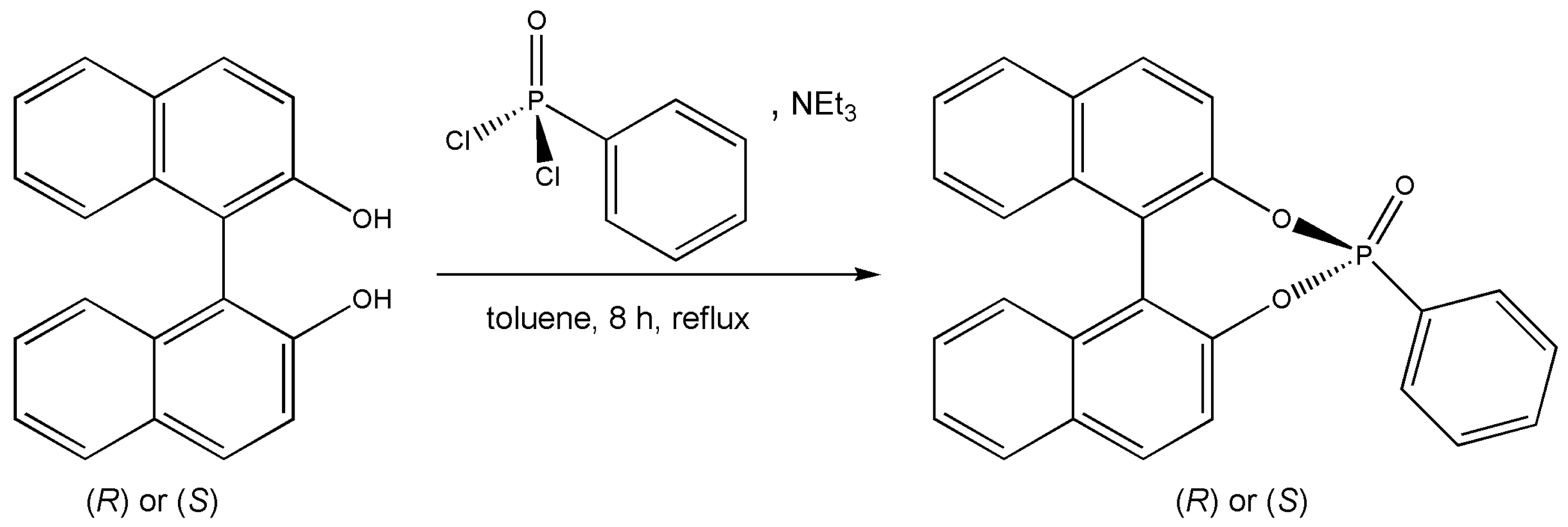
3.2. Synthesis of 4-Phenyldinaphtho[2,1-d:1′,2′-f][1,3,2]dioxaphosphepine 4-Oxide, O=PPh(BINOL)
3.3. Synthesis of [MX2{O=PPh(BINOL)}2] (M = Zn, X = Br; M = Mn, X = Cl, Br)
3.4. Optical Measurements
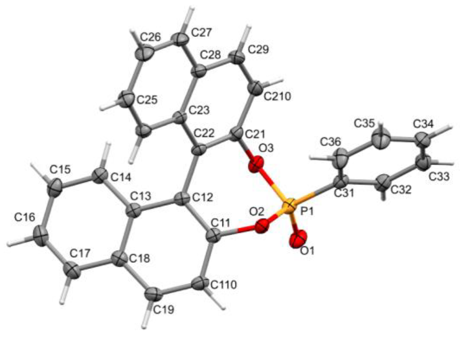
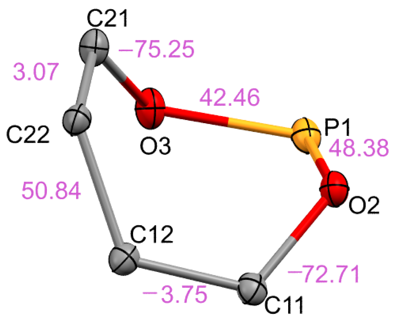
3.5. Crystal Structure Determination
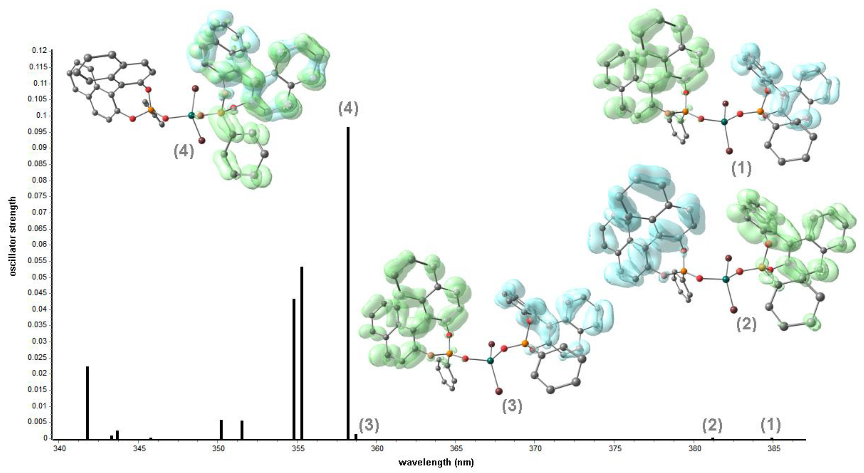
3.6. Computational Details
4. Conclusions
Supplementary Materials
Author Contributions
Funding
Data Availability Statement
Acknowledgments
Conflicts of Interest
References
- Brunel, J.M. BINOL: A Versatile Chiral Reagent. Chem. Rev. 2005, 105, 857–898. [Google Scholar] [CrossRef] [PubMed]
- Chen, Y.; Yekta, S.; Yudin, A.K. Modified BINOL Ligands in Asymmetric Catalysis. Chem. Rev. 2003, 103, 3155–3212. [Google Scholar] [CrossRef] [PubMed]
- Parmar, D.; Sugiono, E.; Raja, S.; Rueping, M. Complete Field Guide to Asymmetric BINOL-Phosphate Derived Brønsted Acid and Metal Catalysis: History and Classification by Mode of Activation; Brønsted Acidity, Hydrogen Bonding, Ion Pairing, and Metal Phosphates. Chem. Rev. 2014, 114, 9047–9153. [Google Scholar] [CrossRef] [PubMed]
- Aspinall, H.C. Chiral Lanthanide Complexes: Coordination Chemistry and Applications. Chem. Rev. 2002, 102, 1807–1850. [Google Scholar] [CrossRef] [PubMed]
- Van Leeuwen, P.W.N.M.; Kamer, P.C.J.; Claver, C.; Pàmies, O.; Diéguez, M. Phosphite-Containing Ligands for Asymmetric Catalysis. Chem. Rev. 2011, 111, 2077–2118. [Google Scholar] [CrossRef]
- Kshatriya, R. Recent Advancement in H8–BINOL Catalyzed Asymmetric Methodologies. ACS Omega 2023, 8, 17381–17406. [Google Scholar] [CrossRef]
- Da Silva, E.M.; Vidal, H.D.A.; Januário, M.A.P.; Corrêa, A.G. Advances in the Asymmetric Synthesis of BINOL Derivatives. Molecules 2023, 28, 12. [Google Scholar] [CrossRef]
- Jurczak, J.; Niedbała, P.; Tyszka-Gumkowska, A. The Synthesis and Application of BINOL Derivatives as Effective Building Blocks for Catalysts Employed in Enantioselective Synthesis. In Chiral Building Blocks in Asymmetric Synthesis: Synthesis and Applications; Wojaczyńska, E., Wojaczyński, J., Eds.; Wiley-VCH: Weinheim, Germany, 2022; pp. 523–549. [Google Scholar] [CrossRef]
- Labelle, A.; Arndtsen, B.A. Chiral BINOL-based borate counterions: From cautionary tale on anion stability to enantioselective Cu-catalyzed cyclopropanation. Chem. Commun. 2023, 59, 728–731. [Google Scholar] [CrossRef]
- Solinas, M.; Meadows, R.E.; Wilson, C.; Blake, A.J.; Woodward, S. Efficient Synthesis of 2-Methyl Derivatives of 1,1′-Bi(2-naphthol) and 1,1′-Bi(2-phenols). Eur. J. Org. Chem. 2007, 2007, 1613–1623. [Google Scholar] [CrossRef]
- Hatano, M.; Yamakawa, K.; Kawai, T.; Horibe, T.; Ishihara, K. Enantioselective Cyanosilylation of Ketones with Lithium(I) Dicyanotrimethylsilicate(IV) Catalyzed by a Chiral Lithium(I) Phosphoryl Phenoxide. Angew. Chem. Int. Ed. 2016, 55, 4021–4025. [Google Scholar] [CrossRef]
- Schlemminger, I.; Lutzen, A.; Willecke, A.; Maison, W.; Koch, R.; Saak, W.; Martens, J. Highly diastereoselective hydrophosphonylation of cyclic imines using BINOL as source of chirality. Tetrahedron Lett. 2000, 41, 7285–7288. [Google Scholar] [CrossRef]
- Teichert, J.F.; Feringa, B.L. Phosphoramidites: Privileged Ligands in Asymmetric Catalysis. Angew. Chem. Int. Ed. 2010, 49, 2486–2528. [Google Scholar] [CrossRef] [PubMed]
- Ma, H.-C.; Sun, Y.-N.; Chen, G.-J.; Dong, Y.-B. A BINOL-phosphoric acid and metalloporphyrin derived chiral covalent organic framework for enantioselective α-benzylation of aldehydes. Chem. Sci. 2022, 13, 1906–1911. [Google Scholar] [CrossRef] [PubMed]
- Zhang, Y.; Yu, W.; Li, H.; Zheng, W.; Cheng, Y. Induced CPL-Active Materials Based on Chiral Supramolecular Co-Assemblies. Chem. Eur. J. 2023, 29, e202204039. [Google Scholar] [CrossRef] [PubMed]
- Feng, H.; Pu, J.; Wang, S.; Jiang, S.; Yang, W.; Cao, D.; Feng, Y.-S. High solid-state CPL active materials based on chiral BINOL-dicyanodistyrylbenzene. Dye. Pigment. 2023, 217, 111422. [Google Scholar] [CrossRef]
- Tauchi, D.; Koida, T.; Nojima, Y.; Hasegawa, M.; Mazaki, Y.; Inagaki, A.; Sugiura, K.-I.; Nagaya, Y.; Tsubaki, K.; Shiga, T.; et al. Aggregation-induced circularly polarized phosphorescence of Pt(II) complexes with an axially chiral BINOL ligand. Chem. Commun. 2023, 59, 4004–4007. [Google Scholar] [CrossRef] [PubMed]
- Liu, C.; Yuan, C.; Shi, G.; Jia, K.; Liu, J.; Wang, K.-P.; Chen, S.; Hu, Z.-Q. Chiral binol-[4]helicene hybrids: Strong solid-state organic emitters with aggregation-enhanced emission and chiroptical properties. Dye. Pigment. 2023, 210, 110992. [Google Scholar] [CrossRef]
- Valverde-González, A.; Borrallo-Aniceto, M.C.; Pintado-Sierra, M.; Sánchez, F.; Arnanz, A.; Boronat, M.; Iglesias, M. BINOL-Containing Chiral Porous Polymers as Platforms for Enantiorecognition. ACS Appl. Mater. Interfaces 2022, 14, 53936–53946. [Google Scholar] [CrossRef]
- Chaudhary, P.; Yadav, G.D.; Singh, S. A simple protocol for determination of enantiopurity of amines using BINOL derivatives as chiral solvating agents via 1H- and 19F-NMR spectroscopic analysis. RSC Adv. 2022, 12, 25457–25464. [Google Scholar] [CrossRef]
- Yu, F.; Chen, Y.; Jiang, H.; Wang, X. Recent advances of BINOL-based sensors for enantioselective fluorescence recognition. Analyst 2020, 145, 6769–6812. [Google Scholar] [CrossRef]
- Van Vliet, S.; Alachouzos, G.; de Vries, F.; Pfeifer, L.; Feringa, B.L. Visible light activated BINOL-derived chiroptical switches based on boron integrated hydrazone complexes. Chem. Sci. 2022, 13, 9713–9718. [Google Scholar] [CrossRef] [PubMed]
- Bull, E.O.J.; Naidu, M.S.R.; Nagaraju, C. In search of new organophosphorus pesticides and insecticides. I: Synthesis and anticholinesterase properties of 4-(substituted phenoxy)-dinaphtho[2,1-d:1′,2′-f] [1,3,2]dioxaphosphepin-4-oxides. Indian J. Chem. 1990, 29B, 688–690. [Google Scholar] [CrossRef]
- Yang, B.; Yue, H.; Shi, D.; Duan, B. An Arylphosphonate Compound and Preparation Method Thereof. CN116425794A, 14 July 2023. [Google Scholar]
- Maekawa, Y.; Kuwabara, K.; Sugiyama, A.; Iwata, K.; Maruyama, T.; Murai, T. Synthesis of P-Stereogenic Phosphinates via an Axis-to-Center Chirality Transfer by the Reaction of Phosphonates Having a Binaphthyloxy Group with Grignard Reagents. Chem. Lett. 2017, 46, 1068–1071. [Google Scholar] [CrossRef]
- Tang, Y.-Y.; Wang, Z.-X.; Li, P.-F.; You, Y.-M.; Stroppa, A.; Xiong, R.-G. Brilliant triboluminescence in a potential organic–inorganic hybrid ferroelectric: (Ph3PO)2MnBr2. Inorg. Chem. Front. 2017, 4, 154–159. [Google Scholar] [CrossRef]
- Huang, X.; Qin, Y.; She, P.; Meng, H.; Liu, S.; Zhao, Q. Functionalized triphenylphosphine oxide-based manganese(II) complexes for luminescent printing. Dalton Trans. 2021, 50, 8831–8836. [Google Scholar] [CrossRef] [PubMed]
- Chen, J.; Zhang, Q.; Zheng, F.-K.; Liu, Z.-F.; Wang, S.-H.; Wu, A.-Q.; Guo, G.-C. Intense photo- and tribo-luminescence of three tetrahedral manganese(II) dihalides with chelating bidentate phosphine oxide ligand. Dalton Trans. 2015, 44, 3289–3294. [Google Scholar] [CrossRef] [PubMed]
- Artem’ev, A.V.; Davydova, M.P.; Berezin, A.S.; Sukhikh, T.S.; Samsonenko, D.G. Photo- and triboluminescent robust 1D polymers made of Mn(II) halides and meta-carborane based bis(phosphine oxide). Inorg. Chem. Front. 2021, 8, 2261–2270. [Google Scholar] [CrossRef]
- Artem’ev, A.V.; Davydova, M.P.; Rakhmanova, M.I.; Bagryanskaya, I.Y.; Pishchur, D.P. A family of Mn(II) complexes exhibiting strong photo- and triboluminescence as well as polymorphic luminescence. Inorg. Chem. Front. 2021, 8, 3767–3774. [Google Scholar] [CrossRef]
- Qin, Y.; Tao, P.; Gao, L.; She, P.; Liu, S.; Li, X.; Li, F.; Wang, H.; Zhao, Q.; Miao, Y.; et al. Designing Highly Efficient Phosphorescent Neutral Tetrahedral Manganese(II) Complexes for Organic Light-Emitting Diodes. Adv. Opt. Mater. 2019, 7, 1801160. [Google Scholar] [CrossRef]
- Li, L.; Wang, Z.-P.; Tian, G.-R.; Song, X.-Y.; Sun, S.-X. Growth and properties of dichloro bis(triphenylphosphine oxide) zinc(II), a novel nonlinear optical crystal. J. Cryst. Growth 2008, 310, 1202–1205. [Google Scholar] [CrossRef]
- Ferraro, V.; Baggio, F.; Castro, J.; Bortoluzzi, M. Green Phosphorescent Zn(II) Halide Complexes with N,N,N′,N′-tetramethyl-P-indol-1-ylphosphonic Diamide as Ligand. Eur. J. Inorg. Chem. 2022, 2022, e202200119. [Google Scholar] [CrossRef]
- Bortoluzzi, M.; Castro, J.; Di Vera, A.; Palù, A.; Ferraro, V. Manganese(II) bromo- and iodo-complexes with phosphoramidate and phosphonate ligands: Synthesis, characterization and photoluminescence. New J. Chem. 2021, 45, 12871–12878. [Google Scholar] [CrossRef]
- Bortoluzzi, M.; Gobbo, A. 1,3-Dimethyl-2-phenyl-1,3-diazaphospholidine-2-oxide as ligand for the preparation of luminescent lanthanide complexes. J. Coord. Chem. 2019, 72, 1524–1536. [Google Scholar] [CrossRef]
- Bortoluzzi, M.; Castro, J.; Gobbo, A.; Ferraro, V.; Pietrobon, L.; Antoniutti, S. Tetrahedral photoluminescent manganese(II) halide complexes with 1,3-dimethyl-2-phenyl-1,3-diazaphospholidine-2-oxide as a ligand. New J. Chem. 2020, 44, 571–579. [Google Scholar] [CrossRef]
- Ferraro, V.; Castro, J.; Agostinis, L.; Bortoluzzi, M. Dual-emitting Mn(II) and Zn(II) halide complexes with 9,10-dihydro-9-oxa-10-phosphaphenanthrene-10-oxide as ligand. Inorg. Chim. Acta 2023, 545, 121285. [Google Scholar] [CrossRef]
- Parsons, S. Determination of absolute configuration using X-ray diffraction. Tetrahedron Asymmetry 2017, 28, 1304–1313. [Google Scholar] [CrossRef]
- Tani, K.; Yamagata, T.; Nagata, K. (±)8-Phenyldinaphtho[2,1-d:1′,2′-f][1,3,2]dioxaphosphepine. Acta Crystallogr. 1994, C50, 1274–1276. [Google Scholar] [CrossRef]
- Pérez, J.; García, J.; Pérez, E.; Serrano, J.L.; Kessler, M. Type conformations and pseudorotation interconversion path-way between conformations: A tool to study medium size (5–9 atoms) rings. J. Mol. Struct. 2012, 1027, 186–199. [Google Scholar] [CrossRef]
- Sinha, S.P.; Pallanen, T.T.; Pakkanen, T.A.; Niinistö, L. Preparation, spectral properties and the crystal structure of the pentacoordinated trichlorobis(hexamethylphosphoramide)-indium (III) complex. Polyhedron 1982, 1, 355–359. [Google Scholar] [CrossRef]
- Liu, Z.; Lu, T.; Chen, Q. An sp-hybridized all-carboatomic ring, cyclo[18]carbon: Electronic structure, electronic spectrum, and optical nonlinearity. Carbon 2020, 165, 461–467. [Google Scholar] [CrossRef]
- Qin, Y.; She, P.; Huang, X.; Huang, W.; Zhao, Q. Luminescent manganese(II) complexes: Synthesis, properties and optoelectronic applications. Coord. Chem. Rev. 2020, 416, 213331. [Google Scholar] [CrossRef]
- Tao, P.; Liu, S.-J.; Wong, W.-J. Phosphorescent manganese(II) complexes and their emerging applications. Adv. Opt. Mater. 2020, 8, 2000985. [Google Scholar] [CrossRef]
- Wrighton, M.; Ginley, D. Excited state decay of tetrahalomanganese(II) complexes. Chem. Phys. 1974, 4, 295–299. [Google Scholar] [CrossRef]
- Englman, R.; Jortner, J. The energy gap law for non-radiative decay in large molecules. J. Lumin. 1970, 1–2, 134–142. [Google Scholar] [CrossRef]
- Wang, Z.-X.; Li, P.-F.; Liao, W.-Q.; Tang, Y.; Ye, H.-Y.; Zhang, Y. Structure-Triggered High Quantum Yield Luminescence and Switchable Dielectric Properties in Manganese(II) Based Hybrid Compounds. Chem. Asian J. 2016, 11, 981–985. [Google Scholar] [CrossRef] [PubMed]
- Artem’ev, A.V.; Davydova, M.P.; Berezin, A.S.; Brel, V.K.; Morgalyuk, V.P.; Bagryanskaya, I.; Samsonenko, D.G. Luminescence of the Mn2+ ion in non-Oh and Td coordination environments: The missing case of square pyramid. Dalton Trans. 2019, 48, 16448–16456. [Google Scholar] [CrossRef] [PubMed]
- Wu, Y.; Zhang, X.; Xu, L.-J.; Yang, M.; Chen, Z.-N. Luminescent Vapochromism Due to a Change of the Ligand Field in a One-Dimensional Manganese(II) Coordination Polymer. Inorg. Chem. 2018, 57, 9175–9181. [Google Scholar] [CrossRef] [PubMed]
- Meng, H.; Zhu, W.; Li, F.; Huang, X.; Qin, Y.; Liu, S.; Yang, Y.; Huang, W.; Zhao, Q. Highly emissive and stable five-coordinated manganese(II) complex for X-ray imaging. Laser Photonics Rev. 2021, 15, 2100309. [Google Scholar] [CrossRef]
- Bortoluzzi, M.; Castro, J.; Ferraro, V. Dual emission from Mn(II) complexes with carbazolyl-substituted phosphoramides. Inorg. Chim. Acta 2022, 536, 120896. [Google Scholar] [CrossRef]
- Bortoluzzi, M.; Ferraro, V.; Castro, J. Synthesis and photoluminescence of manganese(II) naphtylphosphonic diamide complexes. Dalton Trans. 2021, 50, 3132–3136. [Google Scholar] [CrossRef]
- Armarego, W.L.F.; Chai, C.L.L. Purification of Laboratory Chemicals, 5th ed.; Butterworth-Heinemann: London, UK, 2003. [Google Scholar]
- Pietrzyk, D.J.; Frank, C.W. Analytical Chemistry, 2nd ed.; Academic Press: New York, NY, USA, 2012. [Google Scholar]
- Bain, G.A.; Berry, J.F. Diamagnetic corrections and Pascal’s constants. J. Chem. Educ. 2008, 85, 532–536. [Google Scholar] [CrossRef]
- APEX3, SMART, SAINT; Bruker AXS Inc.: Madison, WI, USA, 2015.
- McArdle, P. Oscail, a program package for small-molecule single-crystal crystallography with crystal morphology prediction and molecular modelling. J. Appl. Crystallogr. 2017, 50, 320–326. [Google Scholar] [CrossRef]
- Sheldrick, G.M. SHELXT—Integrated space-group and crystal-structure determination. Acta Crystallogr. 2015, A71, 3–8. [Google Scholar] [CrossRef] [PubMed]
- Sheldrick, G.M. Crystal structure refinement with SHELXL. Acta Crystallogr. 2015, C71, 3–8. [Google Scholar] [CrossRef]
- Spek, A.L. checkCIF validation ALERTS: What they mean and how to respond. Acta Crystallogr. 2020, E76, 1–11. [Google Scholar] [CrossRef] [PubMed]
- Ullrich, C.A. Time-Dependent Density Functional Theory; Oxford University Press: Oxford, UK, 2012. [Google Scholar]
- Grimme, S.; Hansen, A.; Ehlert, S.; Mewes, J.-M. r2SCAN-3c: A “Swiss army knife” composite electronic-structure method. J. Chem. Phys. 2021, 154, 064103. [Google Scholar] [CrossRef]
- Furness, J.W.; Kaplan, A.D.; Ning, J.; Perdew, J.P.; Sun, J. Accurate and Numerically Efficient r2SCAN Meta-Generalized Gradient Approximation. J. Phys. Chem. Lett. 2020, 11, 8208–8215. [Google Scholar] [CrossRef] [PubMed]
- Kruse, H.; Grimme, S. A geometrical correction for the inter- and intra-molecular basis set superposition error in Hartree-Fock and density functional theory calculations for large systems. J. Chem. Phys. 2012, 136, 154101. [Google Scholar] [CrossRef]
- Caldeweyher, E.; Bannwarth, C.; Grimme, S. Extension of the D3 dispersion coefficient model. J. Chem. Phys. 2017, 147, 034112. [Google Scholar] [CrossRef]
- Caldeweyher, E.; Ehlert, S.; Hansen, A.; Neugebauer, H.; Spicher, S.; Bannwarth, C.; Grimme, S. A generally applicable atomic-charge dependent London dispersion correction. J. Chem. Phys. 2019, 150, 154122. [Google Scholar] [CrossRef]
- Brandenburg, J.G.; Bannwarth, C.; Hansen, A.; Grimme, S. B97-3c: A revised low-cost variant of the B97-D density functional method. J. Chem. Phys. 2018, 148, 064104. [Google Scholar] [CrossRef] [PubMed]
- Cossi, M.; Rega, N.; Scalmani, G.; Barone, V. Energies, structures, and electronic properties of molecules in solution with the C-PCM solvation model. J. Comput. Chem. 2003, 24, 669–681. [Google Scholar] [CrossRef] [PubMed]
- Barone, V.; Cossi, M. Quantum calculation of molecular energies and energy gradients in solution by a conductor solvent model. J. Phys. Chem. A 1998, 102, 1995–2001. [Google Scholar] [CrossRef]
- Neese, F. The ORCA program system. WIREs Comput. Mol. Sci. 2012, 2, 73–78. [Google Scholar] [CrossRef]
- Neese, F. Software update: The ORCA program system-Version 5.0. WIREs Comput. Mol. Sci. 2022, 12, e1606. [Google Scholar] [CrossRef]
- Lu, T.; Chen, F. Multiwfn: A multifunctional wavefunction analyzer. J. Comput. Chem. 2012, 33, 580–592. [Google Scholar] [CrossRef]


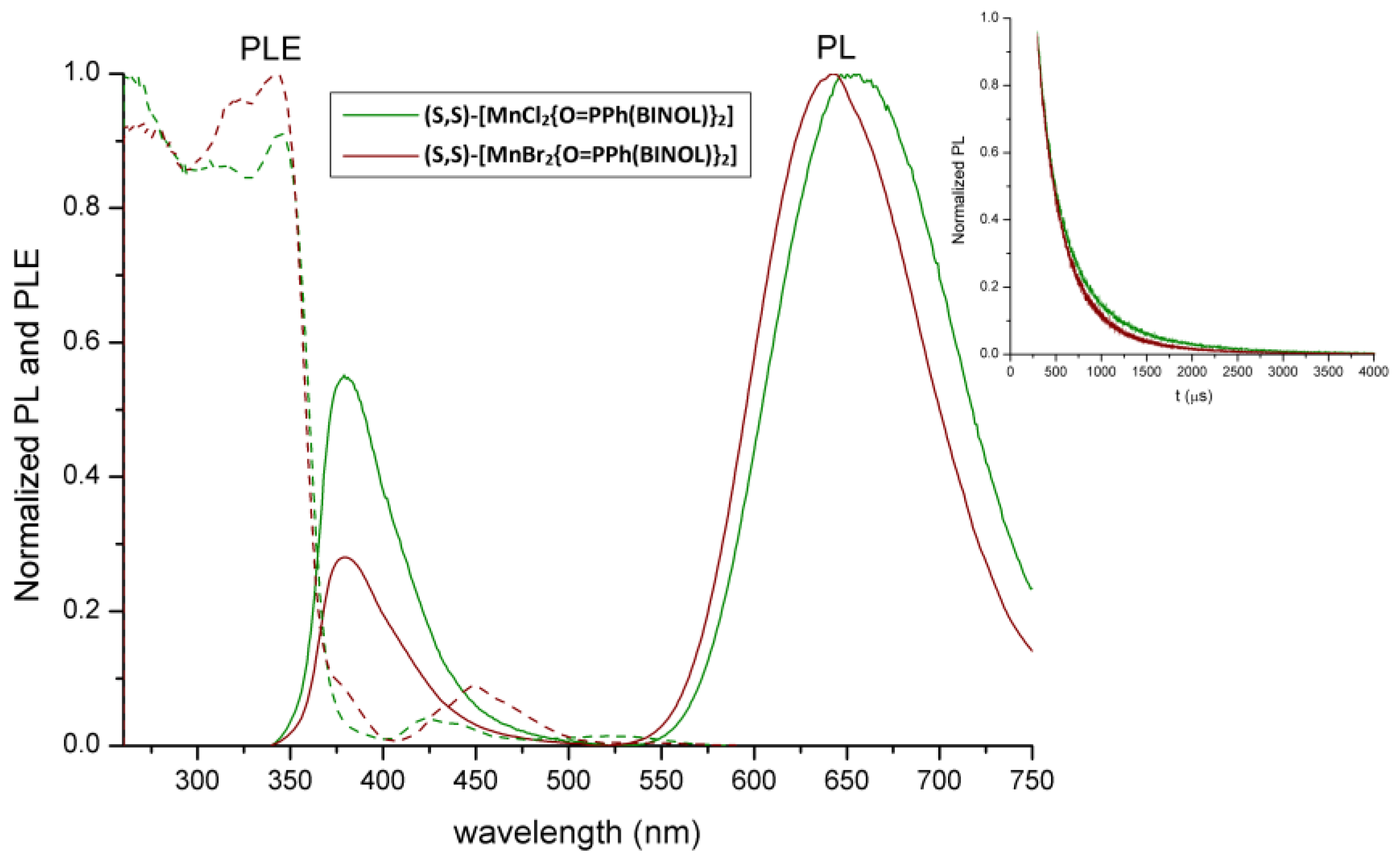
| Complex | PL Max (nm) | PLE (nm) | τ | Φ (%) |
|---|---|---|---|---|
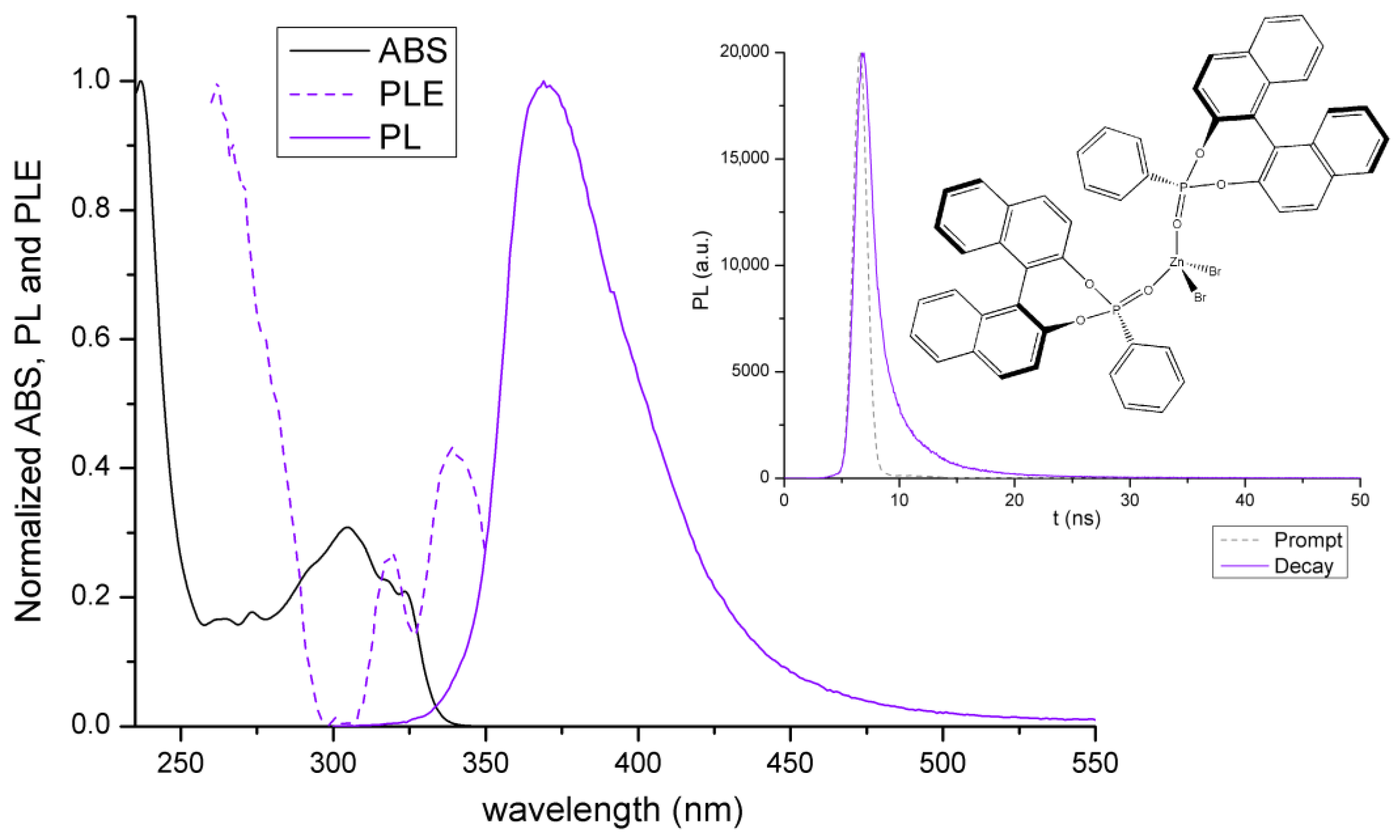
| [ZnBr2{O=PPh(BINOL)}2] | 370 | 320, <300 | 8 ns | 81 |
| [MnCl2{O=PPh(BINOL)}2] | 380, 653 | 410–500, <400 | 220 μs (58%), 700 μs (42%) | 48 |
| [MnBr2{O=PPh(BINOL)}2] | 380, 641 | 410–500, <400 | 216 μs (62%), 530 μs (38%) | 14 |
Disclaimer/Publisher’s Note: The statements, opinions and data contained in all publications are solely those of the individual author(s) and contributor(s) and not of MDPI and/or the editor(s). MDPI and/or the editor(s) disclaim responsibility for any injury to people or property resulting from any ideas, methods, instructions or products referred to in the content. |
© 2024 by the authors. Licensee MDPI, Basel, Switzerland. This article is an open access article distributed under the terms and conditions of the Creative Commons Attribution (CC BY) license (https://creativecommons.org/licenses/by/4.0/).
Share and Cite
Ferraro, V.; Castro, J.; Bortoluzzi, M. Luminescent Behavior of Zn(II) and Mn(II) Halide Derivatives of 4-Phenyldinaphtho[2,1-d:1′,2′-f][1,3,2]dioxaphosphepine 4-Oxide and Single-Crystal X-ray Structure Determination of the Ligand. Molecules 2024, 29, 239. https://doi.org/10.3390/molecules29010239
Ferraro V, Castro J, Bortoluzzi M. Luminescent Behavior of Zn(II) and Mn(II) Halide Derivatives of 4-Phenyldinaphtho[2,1-d:1′,2′-f][1,3,2]dioxaphosphepine 4-Oxide and Single-Crystal X-ray Structure Determination of the Ligand. Molecules. 2024; 29(1):239. https://doi.org/10.3390/molecules29010239
Chicago/Turabian StyleFerraro, Valentina, Jesús Castro, and Marco Bortoluzzi. 2024. "Luminescent Behavior of Zn(II) and Mn(II) Halide Derivatives of 4-Phenyldinaphtho[2,1-d:1′,2′-f][1,3,2]dioxaphosphepine 4-Oxide and Single-Crystal X-ray Structure Determination of the Ligand" Molecules 29, no. 1: 239. https://doi.org/10.3390/molecules29010239