Article

Combined Treatment of Cancer Cells Using Allyl Palladium Complexes Bearing Purine-Based NHC Ligands and Molecules Targeting MicroRNAs miR-221-3p and miR-222-3p: Synergistic Effects on Apoptosis

1
Department of Life Sciences and Biotechnology, University of Ferrara, 44121 Ferrara, Italy
2
Center of Innovative Therapies for Cystic Fibrosis (InnThera4CF), University of Ferrara, 44121 Ferrara, Italy
3
Dipartimento di Scienze Chimiche, Università degli Studi di Padova, 35131 Padova, Italy
4
Dipartimento di Scienze Molecolari e Nanosistemi, University Ca’ Foscari, 30174 Venezia-Mestre, Italy
*
Authors to whom correspondence should be addressed.
These authors contributed equally to this work.
Pharmaceutics 2023, 15(5), 1332; https://doi.org/10.3390/pharmaceutics15051332
Submission received: 21 February 2023 / Revised: 21 April 2023 / Accepted: 21 April 2023 / Published: 24 April 2023
(This article belongs to the Special Issue Development of Novel Tumor-Targeting Nanoparticles)

Abstract

:
Combined treatments employing lower concentrations of different drugs are used and studied to develop new and more effective anticancer therapeutic approaches. The combination therapy could be of great interest in the controlling of cancer. Regarding this, our research group has recently shown that peptide nucleic acids (PNAs) that target miR-221 are very effective and functional in inducing apoptosis of many tumor cells, including glioblastoma and colon cancer cells. Moreover, in a recent paper, we described a series of new palladium allyl complexes showing a strong antiproliferative activity on different tumor cell lines. The present study was aimed to analyze and validate the biological effects of the most active compounds tested, in combination with antagomiRNA molecules targeting two miRNAs, miR-221-3p and miR-222-3p. The obtained results show that a “combination therapy”, produced by combining the antagomiRNAs targeting miR-221-3p, miR-222-3p and the palladium allyl complex 4d, is very effective in inducing apoptosis, supporting the concept that the combination treatment of cancer cells with antagomiRNAs targeting a specific upregulated oncomiRNAs (in this study miR-221-3p and miR-222-3p) and metal-based compounds represents a promising therapeutic strategy to increase the efficacy of the antitumor protocol, reducing side effects at the same time.

1. Introduction

Combination treatments (“combo-therapy”) seem to have a high potential for the development of novel anticancer therapeutic approaches [1,2,3,4,5]. The primary goal of combined treatments is based on the concept and prospect of employing two or more agents in combination in order to have the same biological or therapeutic effect while using lower dosages to reduce recognized adverse effects of the employed agents [1].
In addition, studies have confirmed that an antineoplastic combined therapy can reduce possible acquired resistance [1,2,5]. Therefore, the overall interest in anticancer combined therapy is currently very high [3].
Concerning combined therapeutic approaches, we have recently demonstrated and reported that antitumor derivatives can be combined with molecules targeting specific microRNAs [6,7,8,9], short noncoding RNAs that can operate as gene regulators by directly inhibiting translation or triggering the cleavage of target mRNA transcripts [10,11,12]. MiRNAs may play a role in the pathogenesis of cancer, according to well-established and acknowledged data [13,14]. OncomiRNAs and metastamiRNAs are the terms used to describe miRNAs that are increased in cancer and subsequently cause the downregulation of target tumor suppressor mRNAs [15].
Several studies are available demonstrating synergistic effects of miRNA targeting and use of anticancer drugs [16,17,18]. For instance, Sun et al. developed a core–shell supramolecular nanovector of “chitosome” loaded with Docetaxel (a well-known antineoplastic agent) and with an anti-miRNA molecule against miR-21 to improve chemoresistance in breast tumor [4]. Interestingly, the combination of a conventional chemotherapeutic agent together with the suppression of a specific upregulated oncomiRNA makes cancer cells more sensitive to the treatment, in this case a proposed mechanism is the upregulation of PTEN (an important oncosuppressor that is often inactivated in different types of cancer) which is targeted from miR-21.
In this study, we have employed antagomiRNA molecules targeting miR-221-3p and miR-222-3p on colorectal cancer (CRC) and glioblastoma (GBM) cell lines. MiR-221-3p and miR-222-3p have been selected considering their overexpression in patients with CRC and GBM [19,20,21,22,23]. Moreover, miR-222-3p-targeting decreases cell migration and metastasis using an in vivo GBM [24] or a CRC experimental model systems [25]. Furthermore, the oncogenic role of miR-221-3p was also reported in other types of tumors, such as liver, pancreatic and lung cancer [26,27,28].
Regarding our research activity in this field of investigation, we have designed, synthesized and tested new palladium allyl complexes bearing purine-based N-heterocyclic carbenes derived from the caffeine, theophylline and theobromine scaffolds, characterized by evident cytotoxic and proapoptotic effects on the A2780, Cisplatin-sensitive, and SKOV-3, Cisplatin-resistant, ovarian cancer cell lines [29]. The accurate choice of the supporting ligands is essential for realizing the expected results, and the palladium-allyl fragment was found capable of inducing general cytotoxic effects. The conclusion of this previously published study is that these compounds, and other well-defined organopalladium derivatives, are of great interest for the study of novel anticancer strategies and treatments [29,30,31,32].
The first goal of the present research project was to validate the activity of three different palladium allyl derivatives (4d, 5d and 7a) on cell proliferation of HT29, Ls-174-T and LoVo colon cancer cells, or U251 and T98G glioblastoma cell lines. These three palladium complexes, bearing purine-based carbenes, were designed and synthesized by our research group and previously tested on human ovarian cancer A2780 and SKOV-3 cells [29].
The second aim of our study was to expand the analysis by deepening the activity of these derivatives in order to identify the most bioactive compound, i.e., the best candidate to be used in combination with antagomiRNA molecules against miR-221-3p and miR-222-3p on colon cancer and glioblastoma cancer cells. The selected final endpoint was the ability to induce apoptosis.

2. Materials and Methods

2.1. Chemistry and Reagents

The research group of Prof. Visentin synthesized the palladium compounds 4d, 5d and 7a (Figure 1A); the exploited synthetic procedure has been already reported [29]. All cell cultures required the use of RPMI 1640 medium (cat.no. BE12-702F, Lonza Biosciences, Basel, Switzerland) supplemented with 10% FBS (cat.no. S1400, Biowest, Nuaillè, France) and 100 mg/mL streptomycin and 100 IU/mL penicillin (cat.no. 11074440001, Sigma-Aldrich Merck KGaA). Trypsin-EDTA (cat.no. 59428C, Sigma-Aldrich Merck KGaA, Darmstadt, Hesse, Germany) was utilized to detach the adherent cells, and the Muse® Annexin-V & Dead Cell kit (cat.no. MCH100105, Luminex Corporation, Austin, TX, USA), Muse® Caspase-3/7 kit (cat.no. MCH100108, Luminex Corporation, Austin, TX, USA) was used to perform flow cytometry assays.

2.2. Cell Growth Condition and Cell Lines

Human colon cancer HT29 [33], Ls-174-T [34] and LoVo [35], glioblastoma U251 [36] and T98G [37] cell lines were employed. Cells were seeded in a 12-well plate and maintained at standard conditions (37 °C in a humidified 5% CO2 atmosphere) for 24 h, then treated and incubated for additional 48 h. Following 48 h of treatment, cells were detached from plates with trypsin and counted using a BECKMAN COULTER Z2 cell counter (Beckman, Pasadena, CA, USA). Then, the IC50 values for each compound were calculated. The IC50 represents the 50% inhibitory concentration, which is defined as the compound concentration inhibiting cell proliferation of 50% [7]. The reported IC50 values are average values (±standard deviation) relative to three independent experiments.

2.3. Combined Treatment and Cell Transfection

Cells were seeded in 12-well plates and maintained 24 h at standard conditions (37 °C in a humidified atmosphere 5% CO2) prior to proceeding with the combined treatment. The day after plating, cells were treated and incubated 48 h before proceeding with Apoptosis measurement and RNA extraction; CisPt and compound 4d were employed at sub-optimal concentration (lower than IC50 tested on each cell line). For antimiRNAs transfection, Lipofectamine RNAiMAX (cat.no. 13778075, ThermoFisher, Waltham, MA, USA) was employed following manufacturers’ protocol. In this case, 200 nM antimiR-221-3pp or antimiR-222-3p was diluted in 50 μL of Opti-MEM medium, mixed and incubated 5 min at room temperature with Lipofectamine RNAiMAX in diluted in 50 μL of Opti-MEM prior to treat cells. Cells were treated with CisPt, compound 4d, antimiR-221 or 222 singularly and in combination (CisPt+antimiR-221 or 222 and compound 4d+antimiR-221 or 222); in addition, we put the same amount of Opi-MEM medium in each well and included the proper vehicle controls for single and combined treatment (EtOH, Lipofectamine, or both).

2.4. RNA Extraction

After 48 h of treatment, cells were gently detached with Trypsin-EDTA (cat.no. 59428C, Sigma-Aldrich Merck KGaA, Darmstadt, Hesse, Germany), collected, pelleted in a 1.5 mL tube, and finally lysed with 500 μL of Tri-Reagent (cat.no. 93289, Sigma-Aldrich Merck, Darmstadt, Hesse, Germany). Following a cold 75% ethanol wash, the isolated RNA was kept at −80 °C. Before usage, obtained RNA was vacuum-dried and dissolved in pure and nuclease-free water [9].

2.5. Quantification of miRNAs

MicroRNAs cellular content was investigated using TaqMan MicroRNA Reverse Transcription Kit (cat.no. 43-665-96, Life Technologies, Carlsbad, CA, USA) with RT-qPCR primers and probes that are specific for each target miRNAs tested (listed in Table 1 and purchased from Applied Biosystems, ThermoFisher, Waltham, MA, USA). Each sample was run in duplicate employing TaqMan Universal PCR Master Mix, no AmpErase UNG 2X (cat.no 4324018- ThermoFisher, Waltham, MA, USA) and the CFX96 Touch Real-Time PCR Detection System (BioRad, Hercules, CA, USA). The PCR reaction protocol utilized was the following: 95 °C for 10 min, 95 °C for 15 s, 60 °C for 1 min (last two steps repeated for 50 cycles). Data were obtained and analyzed using Bio-Rad CFX Manager Software (Bio-Rad, Hercules, CA, USA). Relative gene expression was calculated using 2−ΔΔCt method and data normalization was performed using snRNA U6 and hsa-let-7c as endogenous control [9].

2.6. Analysis of Apoptosis

Apoptosis levels were assayed with Guava® Muse® Cell Analyzer instrument and kits (Luminex Corporation, Austin, TX, USA). After 48h of treatment, cell cultures were washed twice with PBS, detached by trypsinization and resuspended in RPMI complete medium. Lastly, 100 μL of cell suspension was incubated for 20 min, protected from light, at room temperature with 100 μL of Muse® Annexin V & 7-AAD (7-aminoactinomycin D) Dead Cell reagent. Cells in the early stage of the apoptotic process (represented in lower right quadrants in the plots) were stained with the Annexin V-PE conjugated; cells in the late phase of apoptosis (upper right quadrants) were stained with Annexin V-PE plus 7-AAD; the necrotic cells were stained only by 7-AAD (upper left quadrants) while live cells showed no staining (lower left quadrants). Following that, samples were obtained using a Guava® Muse® Cell Analyzer (Luminex Corporation, Austin, TX, USA), and data was analyzed using the Luminex Annexin V and Dead Cell Software Module (as previously reported [8]).
Similarly, caspase-3/7 staining was performed with Guava® Muse® Cell Analyzer Caspase-3/7 kit (Luminex Corporation, Austin, TX, USA) following the same protocol of treatment and detachment explained above and following manufacturer’s instruction.

2.7. Statistics

All the reported results are expressed as mean ± standard deviation. The Prism Software was utilized to calculate the comparisons between groups using two-tail paired t-test or ANOVA followed by Dunnet’s multiple comparison. Statistical significance was defined as follows: p < 0.05 (*, significant), p < 0.01 (**, highly significant), p < 0.001 (***, highly significant).

3. Results

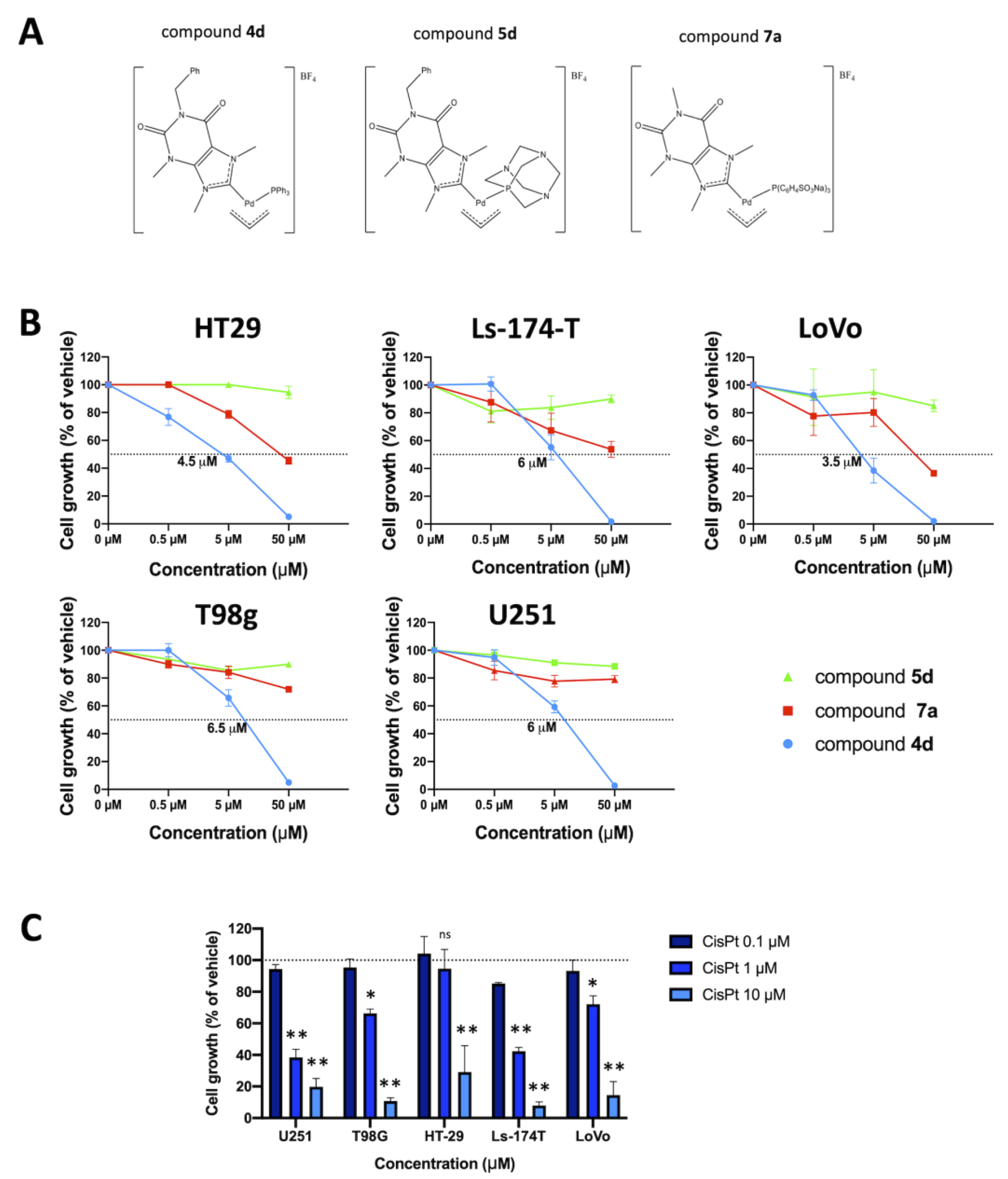
3.1. Structures of the Molecules Employed in This Study and Their Effects on Cell Proliferation

Figure 1A shows the chemical structure of the allyl palladium complexes 4d, 5d and 7a. These compounds have been selected from a panel of palladium allyl complexes synthetized and characterized by some of us in a previously published study [29], which had already shown an excellent antiproliferative activity toward two different lines of ovarian cancer cells. The choice of one phosphine and one N-heterocyclic carbene as ancillary ligands has been made considering the ability of these ligands to stabilize the complexes of transition metals, especially those of 2nd and 3rd transition series. Moreover, the purine-based NHCs should ensure better compatibility with biological environment.
Figure 1B shows the effects of compounds 4d, 5d and 7a on cell proliferation of colon cancer (HT29, Ls-174-T and LoVo) and glioblastoma (U251 and T98G) cell lines. After 72 h of treatment, cell cultures were analyzed to verify the antiproliferative effects of the allyl palladium complexes 4d, 5d and 7a, in comparison with the Cisplatin (CisPt) used at different concentrations. The obtained data indicate that the most effective inhibition of in vitro cell growth is achieved when 4d is used. Figure 1C describes the effects of CisPt on the same cell lines. CisPt was used also in our previous studies and chosen as a positive control, because it is a well-known complex capable of causing apoptosis in various tumor cell lines and, for this reason, still utilized as antineoplastic drug to treat aggressive ovarian and orogastric cancers, although it has many known side effects.
Considering the highest effects of compound 4d, this derivative was selected for the study.
The observed antiproliferative activity of 4d was demonstrated to be associated with activation of apoptosis when the Annexin V assay was employed (Figure 2). Interestingly, induction of apoptosis was detected with high efficiency when compound 4d was assayed using HT29 (Figure 2A), Ls-174-T (Figure 2B), LoVo (Figure 2C), U251 (Figure 2D) and T98G (Figure 2E) cell lines. Moreover, the effects were remarkable when comparison was conducted with CisPt. For instance, when HT29 colon cancer cells were employed, 4d was able to induce 51.60% of late apoptotic cells compared with the 26.05% of late apoptotic cells induced by CisPt (Figure 2A).
In order to verify whether (a) compound 4d synergizes with molecules inhibiting antiapoptotic microRNAs and (b) combined treatments have appreciable effects in T98G drug resistant cells, oligonucleotides able to inhibit miR-221-3p and miR-222-3p were employed. These miRNA targets were selected considering their well-established roles as antiapoptotic regulators [38]. Moreover, the miR-221/miR-222-mRNA networks are well known. For instance, among the potential apoptotic-associated mRNAs, ATG10, CDKN1B/p27, BMF, APAF-1, PTEN, p27(kip1), p57(kip2) and PUMA have been validated as miR-221/miR-222 targets [39,40,41,42,43,44,45,46]. Figure S1, in the “Supplementary Material” section, shows the interactions between miR-221/miR-222 miRNAs and the PUMA 3′-UTR mRNA.

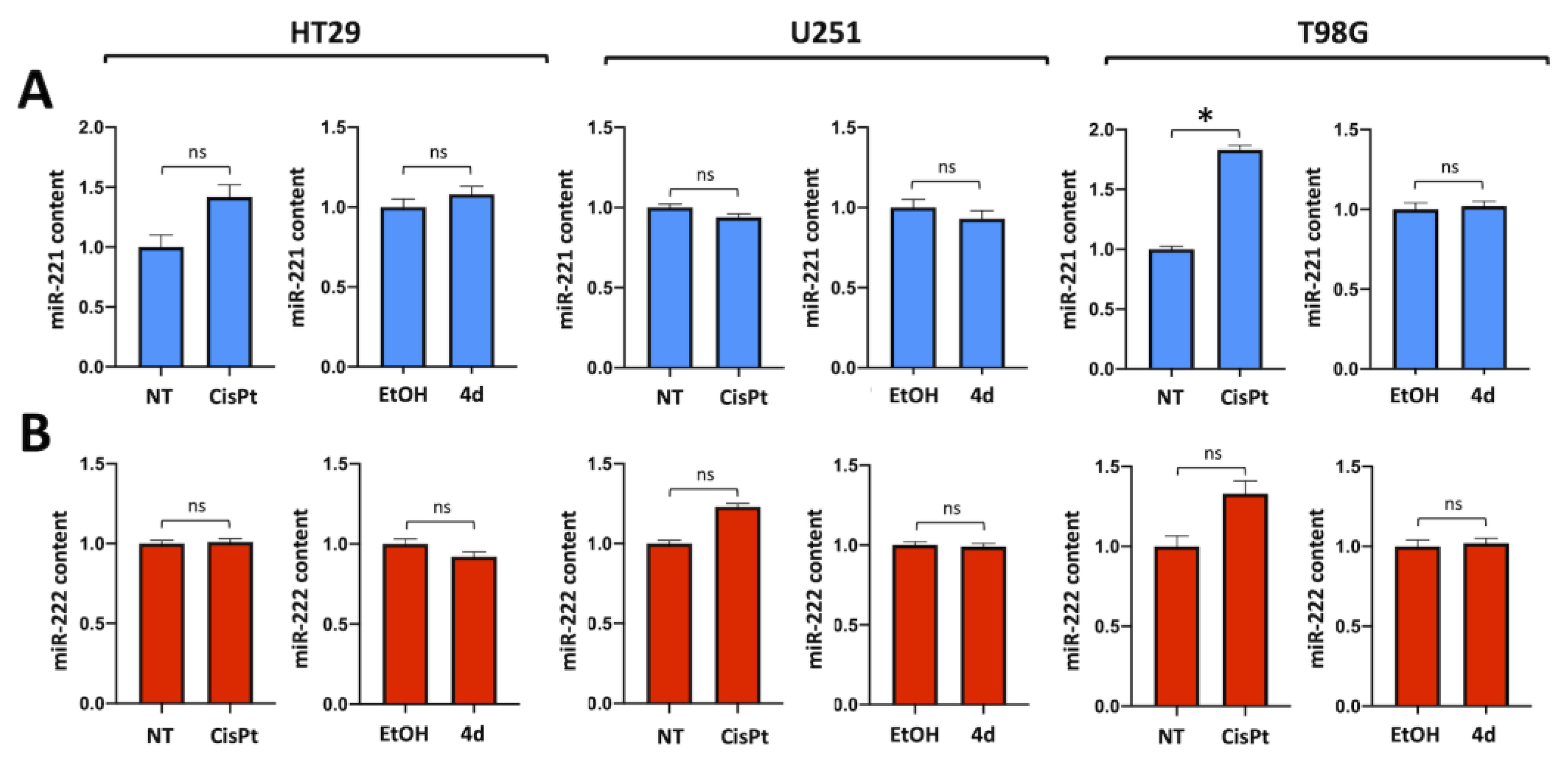
3.2. Compound 4d Does Not Affect Expression of miR-221-3p and miR-222-3p

Compound 4d does not affect miR-221-3p and miR-222-3p in treated HT29 and U251 cells. This experiment, shown in Figure 3, was conducted by exposing, for 48 h, human colon cancer HT29 and human glioblastoma U251 cells to compound 4d or CisPt at IC50 concentration. After this period of cell culture, RNA was isolated, and miR-221-3p (Figure 3A) and miR-222-3p (Figure 3B) were quantified by RT-qPCR. The conclusion of this experiment is that compound 4d does not cause inhibition of the expression of both miR-221-3p and miR-222-3p; similar results were obtained using the T98G temozolomide-resistant glioblastoma cell line.

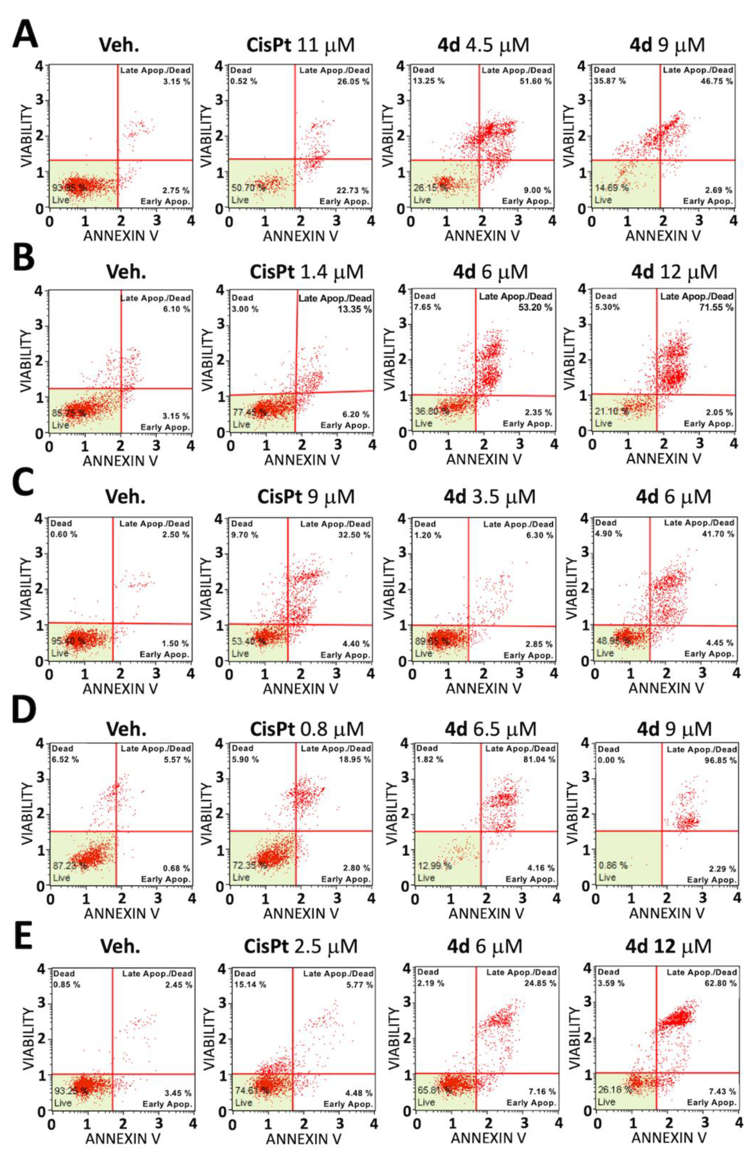
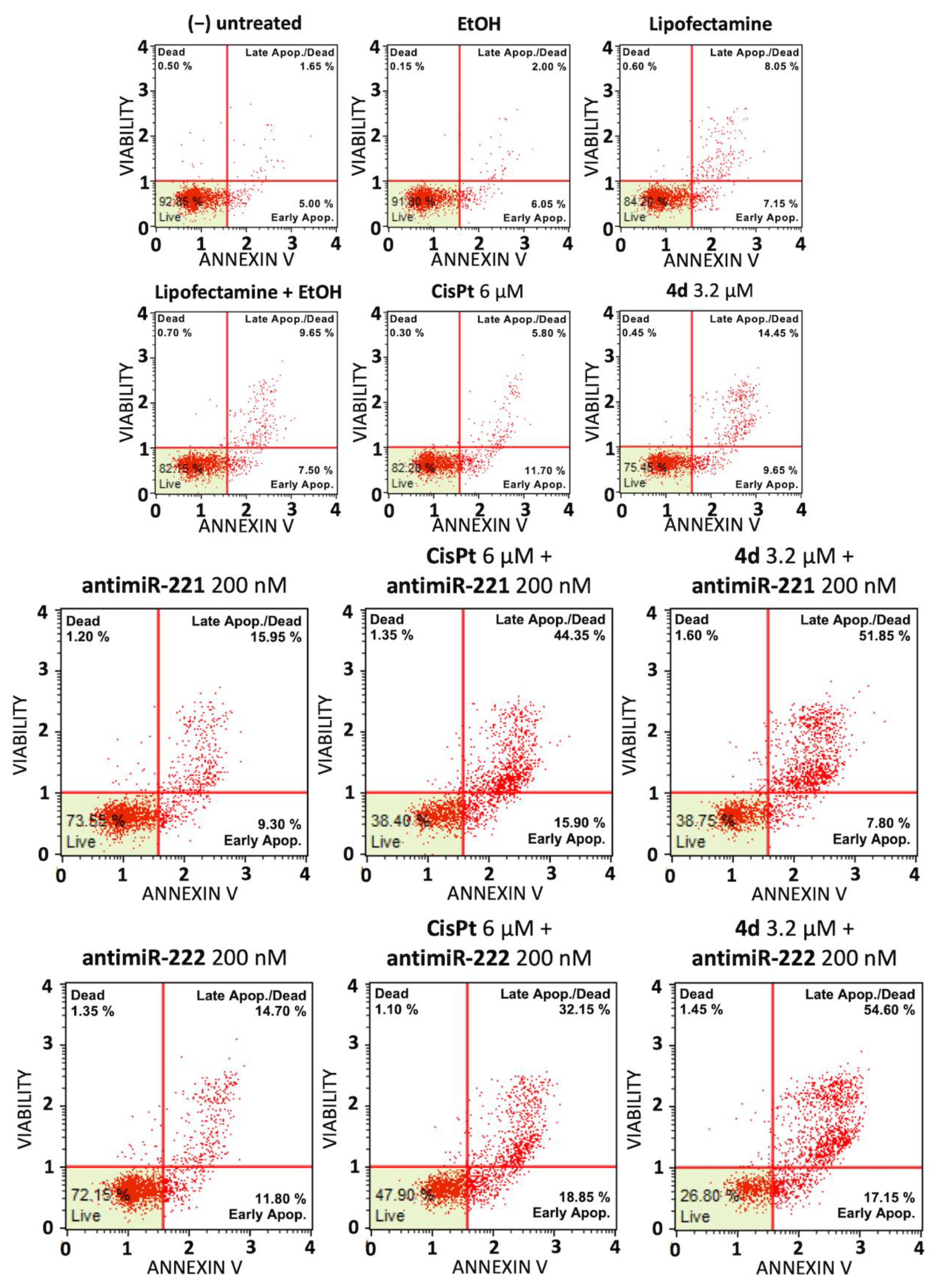
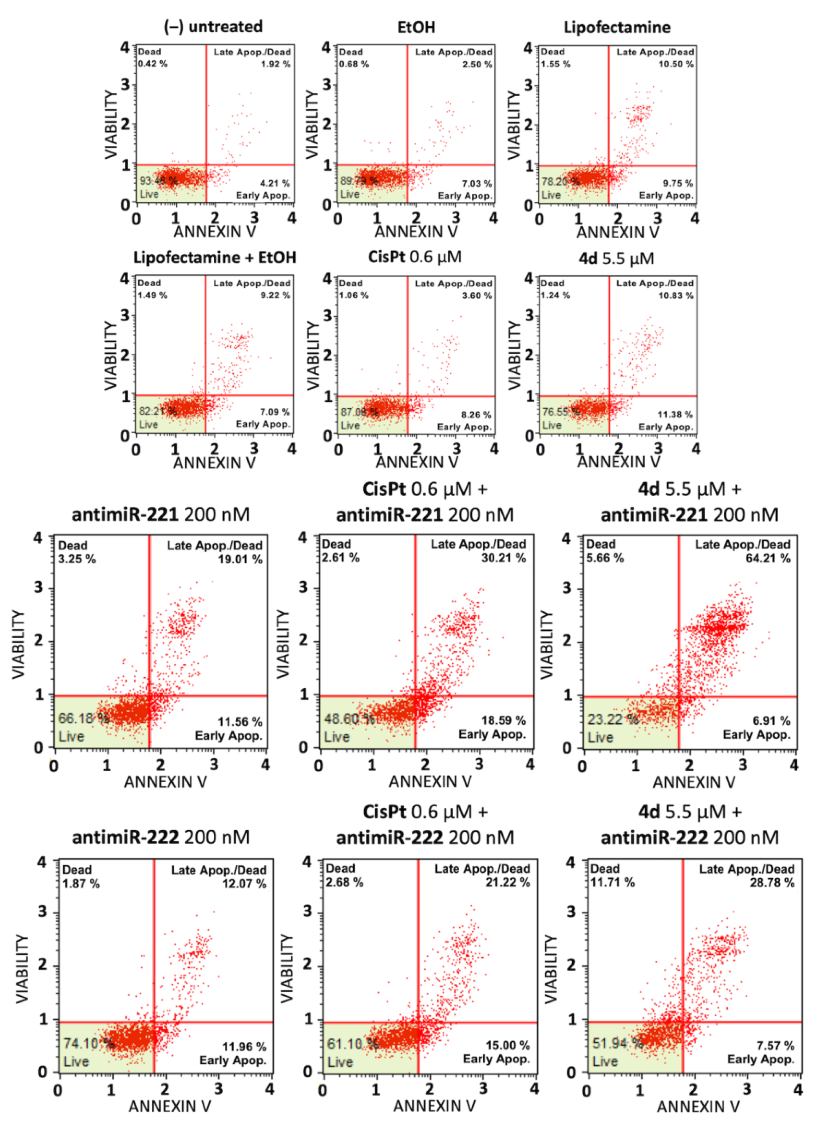
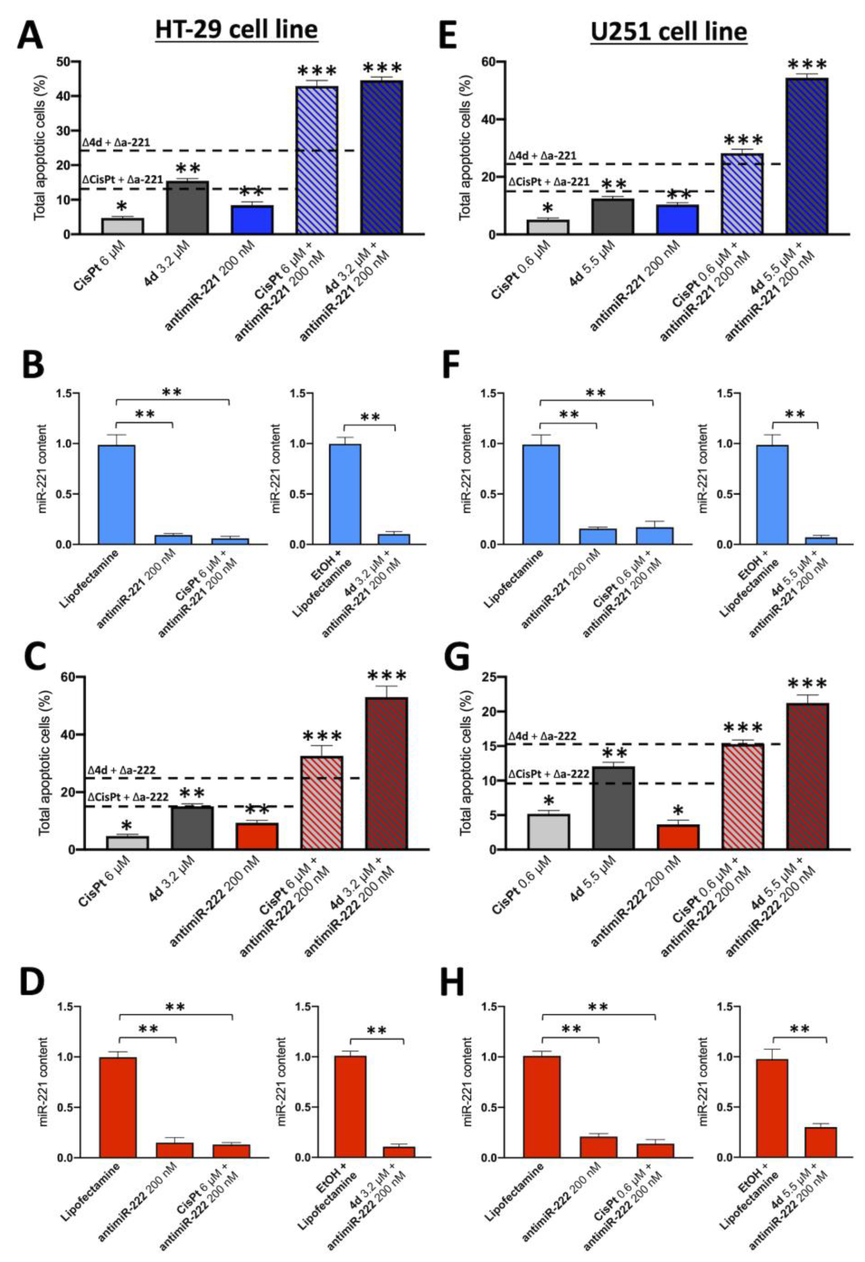
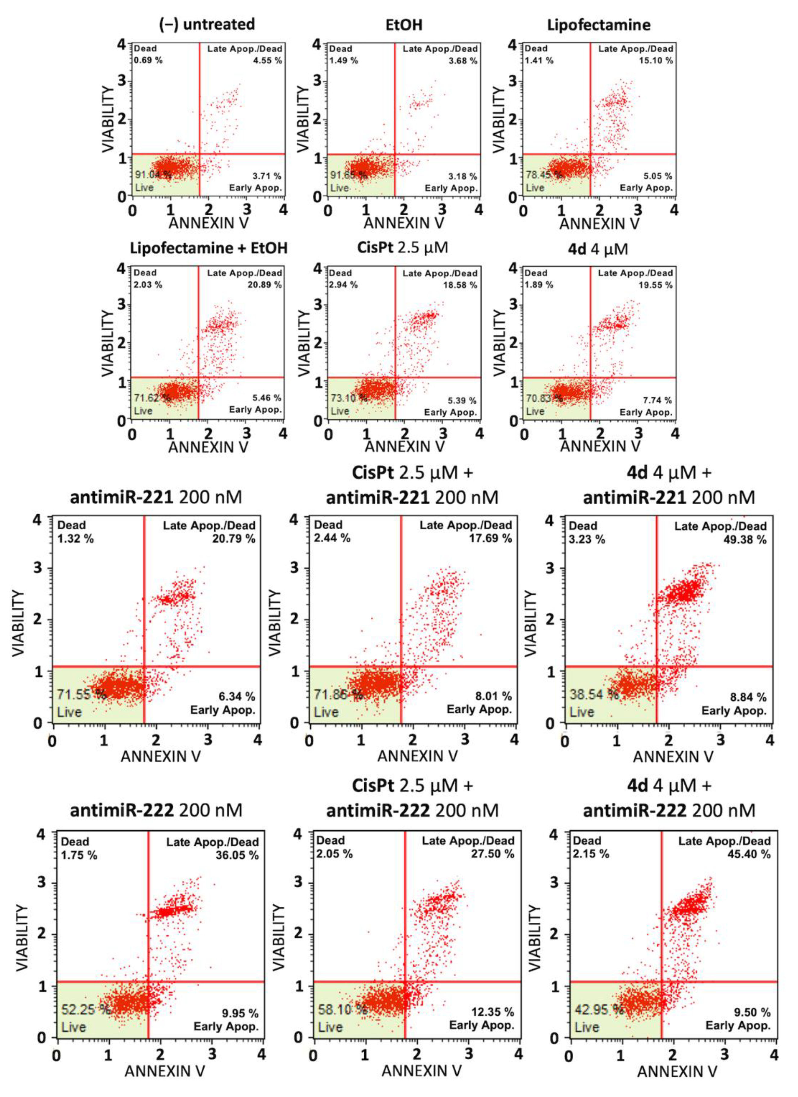
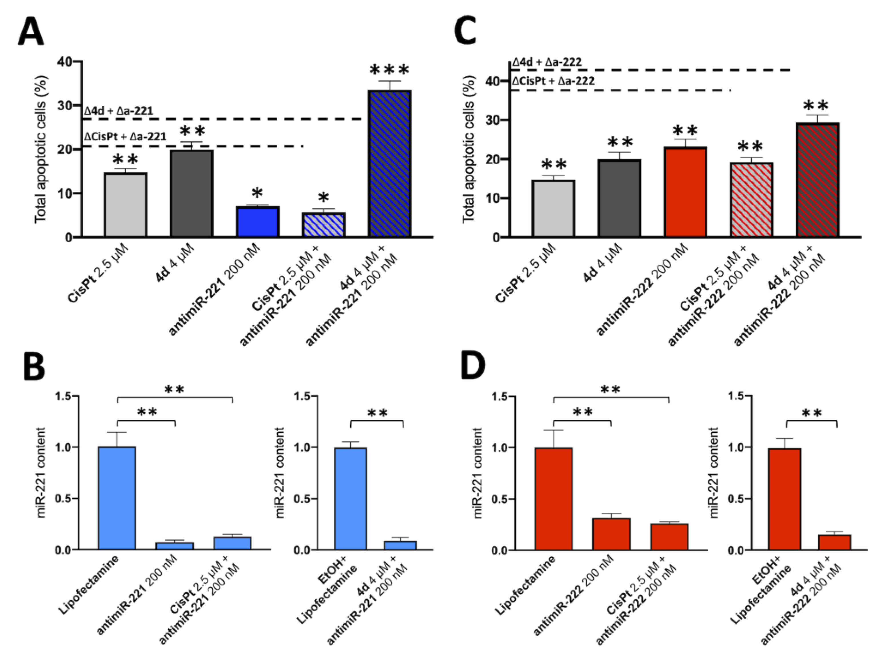
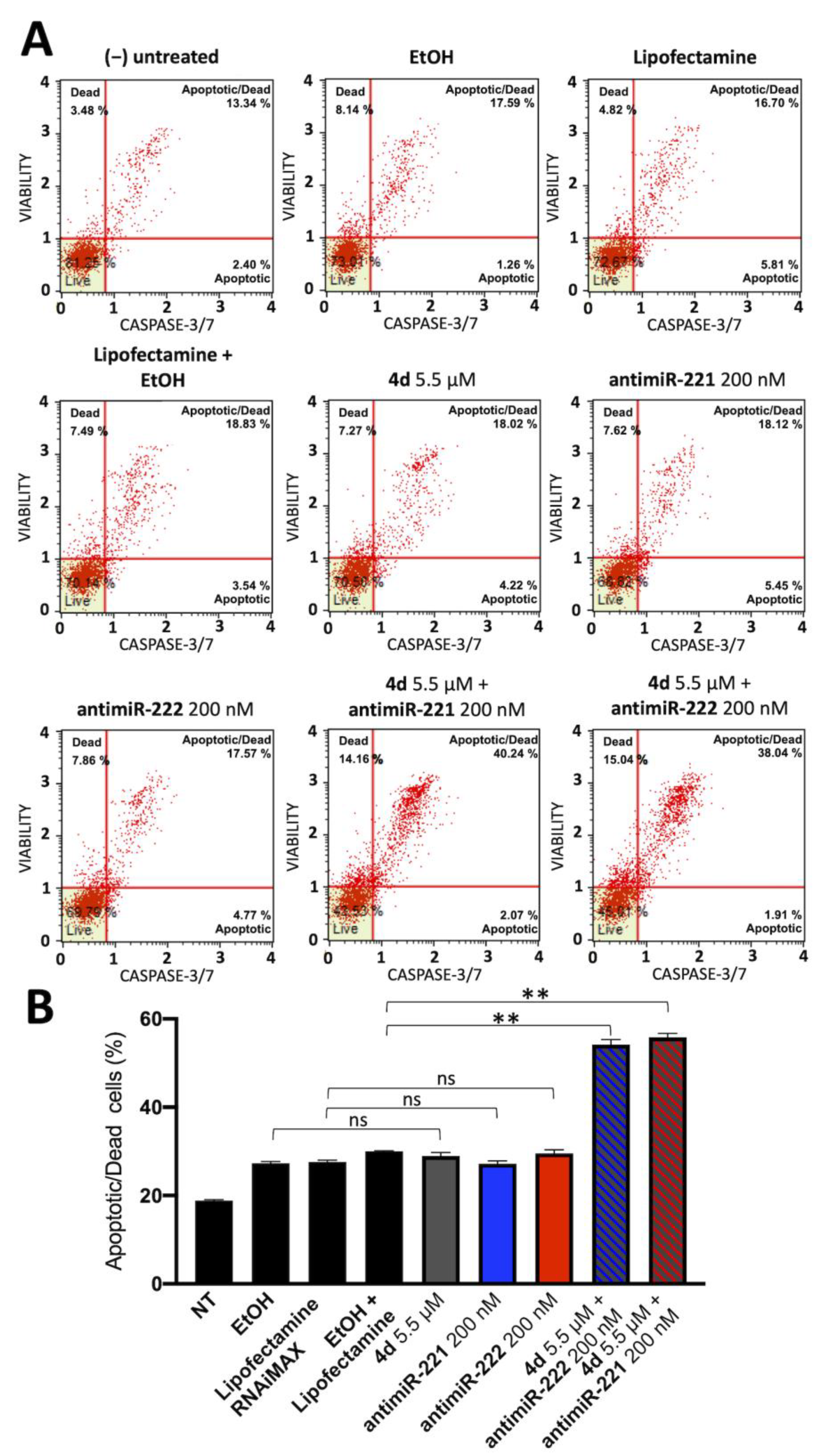
3.3. Co-Treatment of Colon Cancer HT29 and Glioblastoma U251 Cells with 4d and miR-221-3p and miR-222-3p Inhibitors: Effects on Apoptosis Induction

Representative experiments were focused on the effects of single and combined treatments on the induction of apoptosis of HT29 (Figure 4) and U251 (Figure 5) cells. We have comparatively tested the induction of apoptosis (performing Annexin V assay) in the presence of (a) the employed vectors or delivery systems (EtOH, lipofectamine, EtOH+ lipofectamine), (b) the singularly administered 4d, miR-221-3p and miR-222-3p inhibitors and (c) the combined treatments (4d + miR-221-3p inhibitor and 4d + miR-222-3p inhibitor).
The representative experiments shown in Figure 4 and Figure 5 and summarized in Figure 6 conclusively show that the employed vectors or delivery systems (EtOH, lipofectamine, EtOH + lipofectamine) do not induce a significant increase in apoptosis. A second observation is that the singular administration of low concentrations of 4d, CisPt, miR-221-3p inhibitor and miR-222-3p inhibitor stimulates apoptosis. However, the most important conclusion is that the combined treatments based on compound 4d (4d + miR-221-3p inhibitor and 4d + miR-222-3p inhibitor) and on CisPt (CisPt + miR-221-3p inhibitor and CisPt + miR-222-3p inhibitor) lead to an increase in apoptotic cells percentage that was found to be always higher than the sum of the singular treatments. For instance, the sum of the treatment with compound 4d and miR-221-3p inhibitor was 24.11%, while the combined 4d + miR-221 inhibitor was 46.50% (Figure 6A). This very interesting and reproducible result demonstrates a synergism of action. In addition, the combined treatments based on CisPt demonstrated synergistic effects on induction of apoptosis. As expected, down-regulation of miR-221-3p and miR-222-3p relative content was observed in all the treatments in which the miR-221-3p and miR-222-3p inhibitors were used (Figure 6B,D,F,H).

3.4. Cell Apoptosis Study on Glioblastoma Temozolomide-Resistant T98G Cells

To verify whether the synergism of action between compound 4d and the miR-221-3p and miR-222-3p inhibitors occurs also in the temozolomide-resistant T98G cells, the protocol employed on U251 and HT29 cells and described in Figure 4 and Figure 5 was used. The results are shown in Figure 7 and demonstrate that a synergistic action between compound 4d and the miR-221-3p inhibitor is operating also on T98G cells.
The combined treatment, graphed in Figure 8, leads to a clear increase in the apoptosis process which was observed to be higher than the simple sum of the effects obtained using single administration of compound 4d and the miR-221 inhibitor (indicated by the dashed lines). On the contrary no synergistic effects were found with combined treatments based on the miR-222-3p inhibitor and on CisPt.

3.5. Co-Treatment of Glioblastoma U251 Cells with 4d and miR-221-3p and miR-222-3p Inhibitors: Effects on Caspase-3/7 Activation

The effectors in the enzymatic cascade able to activate the apoptotic process are caspases 3 and 7. They are considered biomarkers because they show different phases of the programmed cell death process. To further prove apoptotic protein activation following treatment with compound 4d and miR-221/222 inhibitors, we examined Caspase-3/7 activation by flow cytometry technique as explained in the methods section in U251 glioblastoma cell line. A representative experiment is shown in Figure 9A, while in Figure 9B total apoptotic/dead cells are shown as mean ± standard deviation (n = 3).
While compound 4d and antimiR-221/222 singularly did not show a significant increase in caspase-3/7 activation, the combined treatment with compound 4d and antimiR-221 or antimiR-222 again showed synergistic effects, resulting in a marked increase in caspase-3/7 activation, suggesting that programmed cell death is actively induced following combination treatment.

4. Discussion

Colon cancer (CRC) and Glioblastoma (GBM) patients express high levels of onco-miRNAs, such as miR-221-3p and miR-222-3p, both exerting antiapoptotic effects and promoting malignant progression [21,22,23,24]. The involvement of miR-221-3p and miR-222-3p in CRC has been reported in the studies published by Sanhong et al. [47], demonstrating higher levels of miR-221 and miR-222 in human CRC tissues in respect to healthy colon tissues; this was found to be associated with increased expression of RelA and STAT3 mRNAs; accordingly, interference with the biological activity of miR-221 and miR-222 reduces in vivo growth of colon cancers [47]. The research published by Xu et al. [24] demonstrated that inhibition of both miR-221 and miR-222 significantly lowers proliferation, invasion, migration and angiogenesis of glioblastoma cells in vitro and in vivo. This finding supports the involvement of miR-221-3p in GBM. p-JAK2/JAK2 and p-STAT3/STAT3 pathway activation, levels of various matrix metalloproteinases (including MMP-2 and MMP-9) and levels of vascular endothelial growth factor (VEGF) are all decreased when miR-221-3p and miR-222-3p are inhibited [24].
Considering all these data, we might suggest the miR-221/222 cluster as a pertinent therapeutic target in procedures intended to reduce the invasiveness of GBM and CRC cells during carcinogenesis. An interesting approach that employs combined treatment has been recently proposed by our group, associating compounds interfering with tubulin polymerization and specific antimiRNA molecules targeting upregulated oncomiRNAs (such as miR-221-3p or miR-10b-5p). Combined treatment appears to have synergistic effects on the activation of apoptosis and cell-cycle alterations, demonstrating that this therapeutical strategy can increase the efficacy of anticancer treatments [7,8].
Combined treatments are of clinical interest in the development of anticancer protocols [1,2,3,4,5]. This method limits adverse effects by allowing for the same biological or therapeutic effect to be achieved with two or more medicines administered at lower concentrations [1]. In light of this, combination therapy may be useful in the treatment of glioblastoma, a deadly malignant tumor that requires novel therapeutic choices. In fact, the first-line medicine now employed (temozolomide) is only able to slightly lengthen the life expectancy of GBM patients, who frequently develop chemotherapy resistance. Therefore, there is actually no effective pharmaceutical strategy for glioblastoma, and novel therapeutic protocols are highly needed for clinical validation [48,49,50].
Using inhibitors of miR-221-3p and miR-222-3p along with the palladium allyl derivative 4d, one of the most potent compounds identified by Scattolin et al. [29], we described a novel “combo-therapy” in this study.
The combined treatments of HT29 and U251 cells based on compound 4d (4d + miR-221-3p inhibitor and 4d + miR-222-3p inhibitor) and on CisPt (CisPt + miR-221-3p inhibitor and CisPt + miR-222-3p inhibitor) led to an apoptosis induction that was found to be always higher than the sum of the different treatments. The Combination Index (CI), based on the apoptosis values at different dosages of the drugs, was calculated using the Chou-Talalay method [51]; since the CI was always <1, these obtained results indicate synergistic (rather than additional) effects. As expected, inhibition of miR-221-3p and miR-222-3p was observed in all the treatments in which the miR-221-3p and miR-222-3p inhibitors were used.
Interestingly, when the temozolomide-resistant T98G cell line was used in the experiment, the combined therapy increased apoptosis, in this case more than the combined effects of the single administrations of compound 4d and the miR-221 inhibitor, indicating a synergistic, rather than additive, effect.
In conclusion, the combined use of metal-based anticancer agent (compound 4d) and upregulated “oncomiRNA” inhibitors (miR-221-3p and miR-222-3p in this work) to treat cancer cells is a promising method in the field of developing efficient anticancer therapies, according to our results.

Supplementary Materials

The following supporting information can be downloaded at: https://www.mdpi.com/article/10.3390/pharmaceutics15051332/s1, Figure S1: Structures of miR-221, miR-222 and the 3′-UTR sequence of human PUMA mRNA, showing the interactions between miR-221/miR-222 miRNAs and the PUMA 3′-UTR mRNA. Figure S2: Total apoptotic cells percentage obtained from Annexin-V staining after treatment of HT-29 cell line with CisPt and compound 4d in combination with antimiR-221. Figure S3: Total apoptotic cells percentage obtained from annexin-v staining after treatment of HT-29 cell line with CisPt and compound 4d in combination with antimiR-222. Figure S4: Total apoptotic cells percentage obtained from Annexin-V staining after treatment of U251 cell line with CisPt and compound 4d in combination with antimiR-221. Figure S5: Total apoptotic cells percentage obtained from Annexin-V staining after treatment of U251 cell line with CisPt and compound 4d in combination with antimiR-222. Figure S6: Total apoptotic cells percentage obtained from Annexin-V staining after treatment of T98G cell line with CisPt and compound 4d in combination with an-timiR-221. Figure S7: Total apoptotic cells percentage obtained from Annexin-V staining after treatment of T98G cell line with CisPt and compound 4d in combination with antimiR-222.

Author Contributions

Conceptualization, supervision and experimental design, R.G. and I.L. (Ilaria Lampronti); performing the experiments, M.Z., C.T. and I.L. (Irene Lodi); figures conceptualization and production, M.Z., C.T. and A.F.; writing-review & editing, R.G., I.L. (Ilaria Lampronti), J.G., T.S. and F.V.; project administration and funding acquisition, R.G. and I.L. (Ilaria Lampronti). All authors have read and agreed to the published version of the manuscript.

Funding

This work was supported by the Associazione Italiana per la Ricerca sul Cancro (AIRC) (IG #13575 to R.G.) and FAR (University Fund for Scientific Research to I. Lampronti and A.F.), This study was also supported by the European Union (EU) Horizon 2020 Research and Innovation Programme [GA #633937, project ULTRAsensitive PLAsmonic devices for early CAncer Diagnosis (ULTRAPLACAD) and by the Interuniversity Consortium for the Biotechnology, Italy.

Institutional Review Board Statement

Not applicable.

Informed Consent Statement

Not applicable.

Data Availability Statement

The datasets generated and/or analyzed during the present study are available from the corresponding author upon reasonable request.

Acknowledgments

We wish to thank the “Associazione Tutti per Chiara” (Montagnana, Italy) for supporting Matteo Zurlo with a research fellowship.

Conflicts of Interest

The authors declare no competing interests.

References

  1. Bayat Mokhtari, R.; Homayouni, T.S.; Baluch, N.; Morgatskaya, E.; Kumar, S.; Das, B.; Yeger, H. Combination therapy in combating cancer. Oncotarget 2017, 8, 38022–38043. [Google Scholar] [CrossRef] [PubMed]
  2. Tolcher, A.W.; Mayer, L.D. Improving combination cancer therapy: The CombiPlex® development platform. Future Oncol. 2018, 14, 1317–1332. [Google Scholar] [CrossRef]
  3. Bozic, I.; Reiter, J.G.; Allen, B.; Antal, T.; Chatterjee, K.; Shah, P.; Moon, Y.S.; Yaqubie, A.; Kelly, N.; Le, D.T.; et al. Evolutionary dynamics of cancer in response to targeted combination therapy. eLife 2013, 2, e00747. [Google Scholar] [CrossRef]
  4. Sun, X.; Xu, H.; Huang, T.; Zhang, C.; Wu, J.; Luo, S. Simultaneous delivery of anti-miRNA and docetaxel with supramolecular self-assembled “chitosome” for improving chemosensitivity of triple negative breast cancer cells. Drug Deliv. Trans. Res. 2021, 11, 192–204. [Google Scholar] [CrossRef] [PubMed]
  5. Palmer, A.C.; Sorger, P.K. Combination Cancer Therapy Can Confer Benefit via Patient-to-Patient Variability without Drug Additivity or Synergy. Cell 2017, 171, 1678–1691.e13. [Google Scholar] [CrossRef] [PubMed]
  6. Gasparello, J.; Gambari, L.; Papi, C.; Rozzi, A.; Manicardi, A.; Corradini, R.; Gambari, R.; Finotti, A. High Levels of Apoptosis Are Induced in the Human Colon Cancer HT29 Cell Line by Co-Administration of Sulforaphane and a Peptide Nucleic Acid Targeting miR-15b-5p. Nucleic Acid Ther. 2020, 30, 164–174. [Google Scholar] [CrossRef]
  7. Zurlo, M.; Romagnoli, R.; Oliva, P.; Gasparello, J.; Finotti, A.; Gambari, R. Synergistic effects of the combined treatment of U251 and T98G glioblastoma cells with an anti-tubulin tetrahydrothieno[2,3-c]pyridine derivative and a peptide nucleic acid targeting miR-221-3p. Int. J. Oncol. 2021, 59, 61. [Google Scholar] [CrossRef]
  8. Zurlo, M.; Romagnoli, R.; Oliva, P.; Gasparello, J.; Finotti, A.; Gambari, R. Synergistic Effects of A Combined Treatment of Glioblastoma U251 Cells with An Anti-miR-10b-5p Molecule and An AntiCancer Agent Based on 1-(3′,4′,5′-Trimethoxyphenyl)-2-Aryl-1H-Imidazole Scaffold. Int. J. Mol. Sci. 2022, 23, 5991. [Google Scholar] [CrossRef]
  9. Gasparello, J.; Papi, C.; Zurlo, M.; Gambari, L.; Rozzi, A.; Manicardi, A.; Corradini, R.; Gambari, R.; Finotti, A. Treatment of Human Glioblastoma U251 Cells with Sulforaphane and a Peptide Nucleic Acid (PNA) Targeting miR-15b-5p: Synergistic Effects on Induction of Apoptosis. Molecules 2022, 27, 1299. [Google Scholar] [CrossRef]
  10. Sontheimer, E.J.; Carthew, R.W. Silence from within: Endogenous siRNAs and miRNAs. Cell 2005, 122, 9–12. [Google Scholar] [CrossRef] [PubMed]
  11. Alvarez-Garcia, I.; Miska, E.A. MicroRNA functions in animal development and human disease. Development 2005, 132, 4653–4662. [Google Scholar] [CrossRef] [PubMed]
  12. He, L.; Hannon, G.J. MicroRNAs: Small RNAs with a big role in gene regulation. Nat. Rev. Genet. 2004, 5, 522–531. [Google Scholar] [CrossRef] [PubMed]
  13. Fabbri, M.; Ivan, M.; Cimmino, A.; Negrini, M.; Calin, G.A. Regulatory mechanisms of microRNAs involvement in cancer. Expert Opin. Biol. Ther. 2007, 7, 1009–1019. [Google Scholar] [CrossRef] [PubMed]
  14. Taylor, M.A.; Schiemann, W.P. Therapeutic Opportunities for Targeting microRNAs in Cancer. Mol. Cell. Ther. 2014, 2, 1–13. [Google Scholar] [CrossRef]
  15. Gambari, R.; Brognara, E.; Spandidos, D.A.; Fabbri, E. Targeting oncomiRNAs and mimicking tumor suppressor miRNAs: Νew trends in the development of miRNA therapeutic strategies in oncology (Review). Int. J. Oncol. 2016, 49, 5–32. [Google Scholar] [CrossRef]
  16. Miroshnichenko, S.; Patutina, O. Enhanced Inhibition of Tumorigenesis Using Combinations of miRNA-Targeted Therapeutics. Front. Pharmacol. 2019, 10, 488. [Google Scholar] [CrossRef]
  17. Gajda, E.; Godlewska, M.; Mariak, Z.; Nazaruk, E.; Gawel, D. Combinatory Treatment with miR-7-5p and Drug-Loaded Cubosomes Effectively Impairs Cancer Cells. Int. J. Mol. Sci. 2020, 21, 5039. [Google Scholar] [CrossRef]
  18. Ghasabi, M.; Majidi, J.; Mansoori, B.; Mohammadi, A.; Shomali, N.; Shirafkan, N.; Baghbani, E.; Kazemi, T.; Baradaran, B. The effect of combined miR-200c replacement and cisplatin on apoptosis induction and inhibition of gastric cancer cell line migration. J. Cell. Physiol. 2019, 234, 22581–22592. [Google Scholar] [CrossRef]
  19. Swellam, M.; Ezz El Arab, L.; Al-Posttany, A.S.; Said, S.B. Clinical impact of circulating oncogenic MiRNA-221 and MiRNA-222 in glioblastoma multiform. J. Neurooncol. 2019, 144, 545–551. [Google Scholar] [CrossRef]
  20. Chen, Y.Y.; Ho, H.L.; Lin, S.C.; Ho, T.D.; Hsu, C.Y. Upregulation of miR-125b, miR-181d, and miR-221 Predicts Poor Prognosis in MGMT Promoter-Unmethylated Glioblastoma Patients. Am. J. Clin. Pathol. 2018, 149, 412–417. [Google Scholar] [CrossRef]
  21. Yang, J.K.; Yang, J.P.; Tong, J.; Jing, S.Y.; Fan, B.; Wang, F.; Sun, G.Z.; Jiao, B.H. Exosomal miR-221 targets DNM3 to induce tumor progression and temozolomide resistance in glioblastoma. J. Neurooncol. 2017, 131, 255–265. [Google Scholar] [CrossRef] [PubMed]
  22. Xie, Q.; Yan, Y.; Huang, Z.; Zhong, X.; Huang, L. MicroRNA-221 targeting PI3-K/Akt signaling axis induces cell proliferation and BCNU resistance in human glioblastoma. Neuropathology 2014, 34, 455–464. [Google Scholar] [CrossRef] [PubMed]
  23. Iida, M.; Hazama, S.; Tsunedomi, R.; Tanaka, H.; Takenouchi, H.; Kanekiyo, S.; Tokumitsu, Y.; Tomochika, S.; Tokuhisa, Y.; Sakamoto, K.; et al. Overexpression of miR-221 and miR-222 in the cancer stroma is associated with malignant potential in colorectal cancer. Oncol. Rep. 2018, 40, 1621–1631. [Google Scholar] [CrossRef] [PubMed]
  24. Xu, C.H.; Liu, Y.; Xiao, L.M.; Chen, L.K.; Zheng, S.Y.; Zeng, E.M.; Li, D.H.; Li, Y.P. Silencing microRNA-221/222 cluster suppresses glioblastoma angiogenesis by suppressor of cytokine signaling-3-dependent JAK/STAT pathway. J. Cell. Physiol. 2019, 234, 22272–22284. [Google Scholar] [CrossRef]
  25. Qin, J.; Luo, M. MicroRNA-221 promotes colorectal cancer cell invasion and metastasis by targeting RECK. FEBS Lett. 2014, 588, 99–104. [Google Scholar] [CrossRef]
  26. Dong, Y.; Zhang, N.; Zhao, S.; Chen, X.; Li, F.; Tao, X. miR-221-3p and miR-15b-5p promote cell proliferation and invasion by targeting Axin2 in liver cancer. Oncol. Lett. 2019, 18, 6491–6500. [Google Scholar] [CrossRef] [PubMed]
  27. Li, F.; Xu, J.W.; Wang, L.; Liu, H.; Yan, Y.; Hu, S.Y. MicroRNA-221-3p is up-regulated and serves as a potential biomarker in pancreatic cancer. Artif. Cells Nanomed. Biotechnol. 2018, 46, 482–487. [Google Scholar] [CrossRef]
  28. Yin, G.; Zhang, B.; Li, J. miR-221-3p promotes the cell growth of non-small cell lung cancer by targeting p27. Mol. Med. Rep. 2019, 20, 604–612. [Google Scholar] [CrossRef]
  29. Scattolin, T.; Caligiuri, I.; Canovese, L.; Demitri, N.; Gambari, R.; Lampronti, I.; Rizzolio, F.; Santo, C.; Visentin, F. Synthesis of new allyl palladium complexes bearing purine-based NHC ligands with antiproliferative and proapoptotic activities on human ovarian cancer cell lines. Dalton Trans. 2018, 47, 13616–13630. [Google Scholar] [CrossRef]
  30. Scattolin, T.; Caligiuri, I.; Mouawad, N.; El Boustani, M.; Demitri, N.; Rizzolio, F.; Visentin, F. Synthesis and in-depth studies on the anticancer activity of novel palladacyclopentadienyl complexes stabilized by N-Heterocyclic carbene ligands. Eur. J. Med. Chem. 2019, 179, 325–334. [Google Scholar] [CrossRef] [PubMed]
  31. Scattolin, T.; Giust, S.; Bergamini, P.; Caligiuri, I.; Canovese, L.; Demitri, N.; Gambari, R.; Lampronti, I.; Rizzolio, F.; Visentin, F. Palladacyclopentadienyl complexes bearing purine-based N-heterocyclic carbenes: A new class of promising antiproliferative agents against human ovarian cancer. Appl. Organomet. Chem. 2019, 33, e4902. [Google Scholar] [CrossRef]
  32. Scattolin, T.; Bortolamiol, E.; Visentin, F.; Palazzolo, S.; Caligiuri, I.; Perin, T.; Canzonieri, V.; Demitri, N.; Rizzolio, F.; Togni, A. Palladium(II)-η3 -Allyl Complexes Bearing N-Trifluoromethyl N-Heterocyclic Carbenes: A New Generation of Anticancer Agents that Restrain the Growth of High-Grade Serous Ovarian Cancer Tumoroids. Chemistry 2020, 26, 11868–11876. [Google Scholar] [CrossRef] [PubMed]
  33. Shabahang, M.; Buras, R.R.; Davoodi, F.; Schumaker, L.M.; Nauta, R.J.; Uskokovic, M.R.; Brenner, R.V.; Evans, S.R. Growth inhibition of HT29 human colon cancer cells by analogues of 1,25-dihydroxyvitamin D3. Cancer Res. 1994, 54, 4057–4064. [Google Scholar] [PubMed]
  34. Stein, T.N.; Berger, M.R. Quantification of liver metastases from LS174T human colorectal cancer cells in nude rats by PCR. Anticancer Res. 1999, 19, 3939–3945. [Google Scholar] [PubMed]
  35. Hsu, H.H.; Chen, M.C.; Day, C.H.; Lin, Y.M.; Li, S.Y.; Tu, C.C.; Padma, V.V.; Shih, H.N.; Kuo, W.W.; Huang, C.Y. Thymoquinone suppresses migration of LoVo human colon cancer cells by reducing prostaglandin E2 induced COX-2 activation. World J. Gastroenterol. 2017, 23, 1171–1179. [Google Scholar] [CrossRef] [PubMed]
  36. Cao, X.; Gu, Y.; Jiang, L.; Wang, Y.; Liu, F.; Xu, Y.; Deng, J.; Nan, Y.; Zhang, L.; Ye, J.; et al. A new approach to screening cancer stem cells from the U251 human glioblastoma cell line based on cell growth state. Oncol. Rep. 2013, 29, 1013–1018. [Google Scholar] [CrossRef]
  37. Milani, R.; Brognara, E.; Fabbri, E.; Manicardi, A.; Corradini, R.; Finotti, A.; Gasparello, J.; Borgatti, M.; Cosenza, L.C.; Lampronti, I.; et al. Targeting miR-155-5p and miR-221-3p by peptide nucleic acids induces caspase-3 activation and apoptosis in temozolomide-resistant T98G glioblastoma cells. Int. J. Oncol. 2019, 55, 59–68. [Google Scholar] [CrossRef]
  38. Song, Q.; An, Q.; Niu, B.; Lu, X.; Zhang, N.; Cao, X. Role of miR-221/222 in Tumor Development and the Underlying Mechanism. J. Oncol. 2019, 2019, 7252013. [Google Scholar] [CrossRef]
  39. Shen, H.; Lin, Z.; Shi, H.; Wu, L.; Ma, B.; Li, H.; Yin, B.; Tang, J.; Yu, H.; Yin, X. MiR-221/222 promote migration and invasion, and inhibit autophagy and apoptosis by modulating ATG10 in aggressive papillary thyroid carcinoma. 3 Biotech 2020, 10, 339. [Google Scholar] [CrossRef]
  40. Hu, X.H.; Zhao, Z.X.; Dai, J.; Geng, D.C.; Xu, Y.Z. MicroRNA-221 regulates osteosarcoma cell proliferation, apoptosis, migration, and invasion by targeting CDKN1B/p27. J. Cell. Biochem. 2019, 120, 4665–4674. [Google Scholar] [CrossRef]
  41. Xie, X.; Huang, Y.; Chen, L.; Wang, J. miR-221 regulates proliferation and apoptosis of ovarian cancer cells by targeting BMF. Oncol. Lett. 2018, 16, 6697–6704. [Google Scholar] [CrossRef]
  42. Li, J.; Li, Q.; Huang, H.; Li, Y.; Li, L.; Hou, W.; You, Z. Overexpression of miRNA-221 promotes cell proliferation by targeting the apoptotic protease activating factor-1 and indicates a poor prognosis in ovarian cancer. Int. J. Oncol. 2017, 50, 1087–1096. [Google Scholar] [CrossRef] [PubMed]
  43. Zhou, L.; Jiang, F.; Chen, X.; Liu, Z.; Ouyang, Y.; Zhao, W.; Yu, D. Downregulation of miR-221/222 by a microRNA sponge promotes apoptosis in oral squamous cell carcinoma cells through upregulation of PTEN. Oncol. Lett. 2016, 12, 4419–4426. [Google Scholar] [CrossRef] [PubMed]
  44. Sarkar, S.; Dubaybo, H.; Ali, S.; Goncalves, P.; Kollepara, S.L.; Sethi, S.; Philip, P.A.; Li, Y. Down-regulation of miR-221 inhibits proliferation of pancreatic cancer cells through up-regulation of PTEN, p27(kip1), p57(kip2), and PUMA. Am. J. Cancer Res. 2013, 3, 465–477. [Google Scholar]
  45. Zhang, C.Z.; Zhang, J.X.; Zhang, A.L.; Shi, Z.D.; Han, L.; Jia, Z.F.; Yang, W.D.; Wang, G.X.; Jiang, T.; You, Y.P.; et al. MiR-221 and miR-222 target PUMA to induce cell survival in glioblastoma. Mol. Cancer 2010, 9, 229. [Google Scholar] [CrossRef]
  46. Zhang, C.; Zhang, J.; Zhang, A.; Wang, Y.; Han, L.; You, Y.; Pu, P.; Kang, C. PUMA is a novel target of miR-221/222 in human epithelial cancers. Int. J. Oncol. 2010, 37, 1621–1626. [Google Scholar] [CrossRef]
  47. Liu, S.; Sun, X.; Wang, M.; Hou, Y.; Zhan, Y.; Jiang, Y.; Liu, Z.; Cao, X.; Chen, P.; Liu, Z.; et al. A microRNA 221- and 222-mediated feedback loop maintains constitutive activation of NFκB and STAT3 in colorectal cancer cells. Gastroenterology 2014, 147, 847–859.e11. [Google Scholar] [CrossRef]
  48. Von Neubeck, C.; Seidlitz, A.; Kitzler, H.H.; Beuthien-Baumann, B.; Krause, M. Glioblastoma multiforme: Emerging treatments and stratification markers beyond new drugs. Br. J. Radiol. 2015, 88, 20150354. [Google Scholar] [CrossRef] [PubMed]
  49. Pace, A.; Dirven, L.; Koekkoek, J.A.F.; Golla, H.; Fleming, J.; Rudà, R.; Marosi, C.; Le Rhun, E.; Grant, R.; Oliver, K.; et al. European Association for Neuro-Oncology (EANO) guidelines for palliative care in adults with glioblastoma. Lancet Oncol. 2017, 18, e330–e340. [Google Scholar] [CrossRef]
  50. Anjum, K.; Shagufta, B.I.; Abbas, S.Q.; Patel, S.; Khan, I.; Shah, S.A.A.; Akhter, N.; Hassan, S.S.U. Current status and future therapeutic perspectives of glioblastoma multiforme (GBM) therapy: A review. Biomed. Pharmacother. 2017, 92, 681–689. [Google Scholar] [CrossRef]
  51. Chou, T.-C. Drug combination studies and their synergy quantification using the Chou-Talalay method. Cancer Res. 2010, 70, 440–446. [Google Scholar] [CrossRef] [PubMed]
Figure 1. Chemical structure of molecules employed in this study and their effects on cell proliferation. (A) Chemical structure of the allyl palladium complexes 4d, 5d and 7a. (B) Effects of compounds 4d (blue lines and symbols), 5d (green lines and symbols) and 7a (red lines and symbols) on cell proliferation of different colon cancer (HT29, Ls-174-T and LoVo) and glioblastoma (U251 and T98G) cell lines, after 72 h of incubation; crossing of colored lines with the dotted line represents the IC50 value. (C) Effects of Cisplatin (CisPt) at different concentrations (0.1, 1, 10 μM) on cell proliferation of the same cell lines. Results represent mean ± SD (n = 3). p < 0.05 (*, significant), p < 0.01 (**; highly significant), ns = not significant, in comparison to the negative control (untreated cells), shown by the dotted line (100% cell growth).
Pharmaceutics 15 01332 g001
Figure 2. Induction of apoptosis of compound 4d and Cisplatin (CisPt) in representative experiments. Annexin V/7-AAD assay was employed. HT29 (A), Ls-174-T (B), LoVo (C), U251 (D) and T98G (E) cells were treated with compound 4d at IC50 and IC75 concentration or Cisplatin (IC50) for 48 h and then assayed for apoptosis induction, in comparison with cells treated with vehicle (veh.).
Pharmaceutics 15 01332 g002
Figure 3. Cisplatin (CisPt) and compound 4d do not act downregulating miR221 (A) or miR222 (B) in colorectal HT29 and glioblastomaU251 and T98G cell lines. CisPt and complex 4d treatment normally do not interfere with miR-221/222 expression in tested cell lines. RT-qPCR data were normalized with housekeeping U6 snRNA, cells treated with water-soluble CisPt were compared with untreated cells (NT) while cells treated with 4d, soluble in ethanol, were compared with cells treated with vehicle (EtOH). Results represent mean ± SD (n = 3). p < 0.05 (*, significant), ns = not significant.
Pharmaceutics 15 01332 g003
Figure 4. Induction of apoptosis of combined treatments based on the compound 4d and inhibitors of miR-221-3p and miR-222-3p: colon cancer HT29 cell line. Annexin V assay was employed. HT29 cells were treated for 48 h with the indicated experimental conditions, i.e., with the employed vectors or delivery systems (EtOH, lipofectamine, EtOH + lipofectamine) and the singularly administered 4d and the positive control CisPt (upper panel), with singularly administered miR-221-3p and miR-222-3p inhibitors and with the combined treatments using the miR-221-3p and miR-222-3p inhibitors and 4d or CisPt (lower panel).
Pharmaceutics 15 01332 g004
Figure 5. Induction of apoptosis of combined treatments based on the compound 4d and inhibitors of miR-221-3p and miR-222-3p: glioblastoma U251 cell line. Annexin V assay was employed. U251 cells were treated for 48 h with the indicated experimental conditions, i.e., with the employed vectors or delivery systems (EtOH, lipofectamine, EtOH + lipofectamine) and with the singularly administered 4d and the positive control CisPt (upper panel), with singularly administered miR-221-3p and miR-222-3p inhibitors and with the combined treatments using the miR-221-3p and miR-222-3p inhibitors and 4d or CisPt (lower panel).
Pharmaceutics 15 01332 g005
Figure 6. Summary of the experiments performed on the induction of apoptosis of combined treatments based on the compound 4d and inhibitors of miR-221-3p and miR-222-3p. Experiments performed on HT29 cell line (AD) are displayed on the left, while U251 cells (EH) are displayed on the right; (A,C,E,G): effects on the induction of apoptosis in combined treatments based on miR-221-3p (A,E) and miR-222-3p (C,G) inhibitors; (B,D,F,H): effects of the indicated treatments on accumulation of miR-221-3p (B,F) and miR-222-3p (D,H). The dotted lines of panels (A,C,E,G) represent the sum of the singularly administered compounds, as indicated. Results represent mean ± SD (n = 3). p < 0.05 (*, significant), p < 0.01 (**; highly significant), p < 0.001 (***; highly significant).
Pharmaceutics 15 01332 g006
Figure 7. Induction of apoptosis of combined treatments based on the compound 4d and inhibitors of miR-221-3p and miR-222-3p: glioblastoma T98G cell line. Annexin V assay was employed. T98G cells were treated for 48 h with the indicated experimental conditions, i.e., with the employed vectors or delivery systems (EtOH, lipofectamine, EtOH + lipofectamine) and with the singularly administered 4d and the positive control CisPt (upper panel), with singularly administered miR-221-3p and miR-222-3p inhibitors and with the combined treatments using the miR-221-3p and miR-222-3p inhibitors and 4d or CisPt (lower panel).
Pharmaceutics 15 01332 g007
Figure 8. Summary of the experiments performed on the induction of apoptosis of combined treatments based on the compound 4d and inhibitors of miR-221-3p and miR-222-3p: glioblastoma T98G cells. (A,C): Annexin V assay. (B,D): RT-qPCR analysis of the miR-221-3p and miR-222-3p cellular content. The dotted lines of panel (A,C) represent the sum of the singularly administered compounds, as indicated. Results represent mean ± SD (n = 3). p < 0.05 (*, significant), p < 0.01 (**; highly significant), p < 0.001 (***; highly significant).
Pharmaceutics 15 01332 g008
Figure 9. Summary of the experiments performed on U251 cell line showing caspase-3/7 activation following combined treatments based on the compound 4d and inhibitors of miR-221-3p and miR-222-3p. (A): Representative dot plot obtained by flow cytometry following Caspase-3/7 staining. (B): Histogram representing the mean increase in apoptotic/dead cells, results represent mean ± SD (n = 3). p < 0.01 (**; highly significant), ns = not significant.
Pharmaceutics 15 01332 g009
Table 1. List of assays employed for miRNA detection.
miRNA NameAssay ID
hsa-miR-221-3p
has-miR-222-3p
000524
000525
hsa-snRNA U6
hsa-let-7c-5p
001973
000379
Disclaimer/Publisher’s Note: The statements, opinions and data contained in all publications are solely those of the individual author(s) and contributor(s) and not of MDPI and/or the editor(s). MDPI and/or the editor(s) disclaim responsibility for any injury to people or property resulting from any ideas, methods, instructions or products referred to in the content.

